HDAC1 Antibody
Novus Biologicals, part of Bio-Techne | Catalog # NBP2-54621

Key Product Details
Species Reactivity
Human, Mouse
Applications
Chromatin Immunoprecipitation (ChIP), Chromatin Immunoprecipitation Sequencing, ELISA, Immunocytochemistry/ Immunofluorescence, Knockdown Validated, Protein Array, Western Blot
Label
Unconjugated
Antibody Source
Polyclonal Rabbit IgG
Concentration
Please see the vial label for concentration. If unlisted please contact technical services.
Product Specifications
Immunogen
HDAC1
Clonality
Polyclonal
Host
Rabbit
Isotype
IgG
Scientific Data Images for HDAC1 Antibody
Western Blot: HDAC1 Antibody [NBP2-54621]
Western Blot: HDAC1 Antibody [NBP2-54621] - Whole cell extracts (50 ug) from HeLa cells transfected with HDAC1 siRNA (lane 2) and from an untransfected control (lane 1) were analysed by Western blot using the antibody against HDAC1 diluted 1:1,000 in TBS-Tween containing 5% skimmed milk. The position of the protein of interest is indicated on the right (expected size: 55 kDa); the marker (in kDa) is shown on the left.Immunocytochemistry/ Immunofluorescence: HDAC1 Antibody [NBP2-54621]
Immunocytochemistry/Immunofluorescence: HDAC1 Antibody [NBP2-54621] - HeLa cells were stained with the antibody against HDAC1 and with DAPI. Cells were fixed with 4% formaldehyde for 10' and blocked with PBS/TX-100 containing 5% normal goat serum and 1% BSA. The cells were immunofluorescently labelled with the HDAC1 antibody (left) diluted 1:500 in blocking solution followed by an anti-rabbit antibody conjugated to Alexa488. The middle panel shows staining of the nuclei with DAPI. A merge of the two stainings is shown on the right.
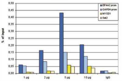
Chromatin Immunoprecipitation: HDAC1 Antibody [NBP2-54621] - ChIP was performed with the antibody against HDAC1 on sheared chromatin from 4,000,000 HeLa cells. An antibody titration consisting of 1, 2, 5 and 10 ug per ChIP experiment was analysed. IgG (2 ug/IP) was used as negative IP control. QPCR was performed with primers specific for the EIF4A2 and GAPDH promoters, used as positive controls, and for the MYOD1 gene and Sat2 satellite repeat, used as negative controls. Figure shows the recovery, expressed as a % of input (the relative amount of immunoprecipitated DNA compared to input DNA after qPCR analysis).
Applications for HDAC1 Antibody
Application
Recommended Usage
Chromatin Immunoprecipitation (ChIP)
2 ug/IP
ELISA
1:4000
Immunocytochemistry/ Immunofluorescence
1:500
Protein Array
1:100000
Western Blot
1:1000
Formulation, Preparation, and Storage
Purification
Affinity purified
Formulation
PBS
Preservative
0.05% Sodium Azide and 0.05% ProClin 300
Concentration
Please see the vial label for concentration. If unlisted please contact technical services.
Shipping
The product is shipped with polar packs. Upon receipt, store it immediately at the temperature recommended below.
Stability & Storage
Store at -20C. Avoid freeze-thaw cycles.
Background: Histone Deacetylase 1/HDAC1
Long Name
Histone Deacetylase 1
Alternate Names
GON-10, HD1, KDAC1, RPD3L1
Gene Symbol
HDAC1
Additional Histone Deacetylase 1/HDAC1 Products
Product Specific Notices for HDAC1 Antibody
This product is for research use only and is not approved for use in humans or in clinical diagnosis. Primary Antibodies are guaranteed for 1 year from date of receipt.
Loading...
Loading...
Loading...
Loading...
![Immunocytochemistry/ Immunofluorescence: HDAC1 Antibody [NBP2-54621] Immunocytochemistry/ Immunofluorescence: HDAC1 Antibody [NBP2-54621]](https://resources.bio-techne.com/images/products/HDAC1-Antibody-Immunofluorescence-NBP2-54621-img0006.jpg)

![Western Blot: HDAC1 Antibody [NBP2-54621] Western Blot: HDAC1 Antibody [NBP2-54621]](https://resources.bio-techne.com/images/products/HDAC1-Antibody-Western-Blot-NBP2-54621-img0003.jpg)
![Western Blot: HDAC1 Antibody [NBP2-54621] Western Blot: HDAC1 Antibody [NBP2-54621]](https://resources.bio-techne.com/images/products/HDAC1-Antibody-Western-Blot-NBP2-54621-img0004.jpg)
![ELISA: HDAC1 Antibody [NBP2-54621] ELISA: HDAC1 Antibody [NBP2-54621]](https://resources.bio-techne.com/images/products/HDAC1-Antibody-ELISA-NBP2-54621-img0002.jpg)
![Protein Array: HDAC1 Antibody [NBP2-54621] Protein Array: HDAC1 Antibody [NBP2-54621]](https://resources.bio-techne.com/images/products/HDAC1-Antibody-Protein-Array-NBP2-54621-img0005.jpg)