Human C1D Antibody
R&D Systems, part of Bio-Techne | Catalog # AF4816

Key Product Details
Species Reactivity
Applications
Label
Antibody Source
Product Specifications
Immunogen
Ala2-Ser141
Accession # Q13901
Specificity
Clonality
Host
Isotype
Scientific Data Images for Human C1D Antibody
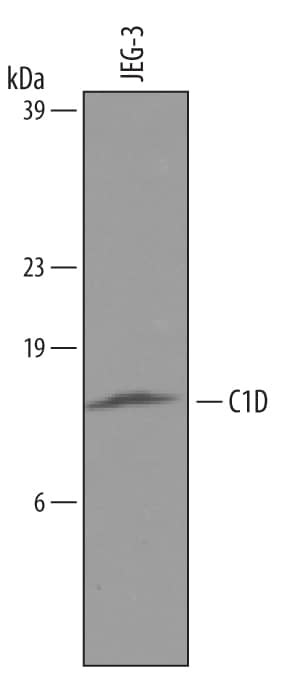
Detection of Human C1D by Western Blot.
Western blot shows lysates of JEG-3 human epithelial choriocarcinoma cell line. PVDF membrane was probed with 2 µg/mL of Goat Anti-Human C1D Antigen Affinity-purified Polyclonal Antibody (Catalog # AF4816) followed by HRP-conjugated Anti-Goat IgG Secondary Antibody (Catalog # HAF019). A specific band was detected for C1D at approximately 16 kDa (as indicated). This experiment was conducted under reducing conditions and using Immunoblot Buffer Group 8.Applications for Human C1D Antibody
Western Blot
Sample: JEG-3 human epithelial choriocarcinoma cell line
Formulation, Preparation, and Storage
Purification
Reconstitution
Formulation
Shipping
Stability & Storage
- 12 months from date of receipt, -20 to -70 °C as supplied.
- 1 month, 2 to 8 °C under sterile conditions after reconstitution.
- 6 months, -20 to -70 °C under sterile conditions after reconstitution.
Background: C1D
C1D (also SUN-CoR) is a 16 kDa member of the C1D family of DNA-binding proteins. It is ubiquitously expressed, activates DNA‑dependent protein kinase, and forms part of a large complex that processes rRNA. C1D exists as both a monomer and noncovalent homodimer, and is known to be phosphorylated. Human C1D is 141 amino acids (aa) in length. It contains two protein interaction sites at aa 50‑100 and 101‑141. There is one potential alternate site at Met54, and a number of single aa polymorphisms. Full-length C1D is 90% and 87% aa identical to mouse and canine C1D, respectively.
Long Name
Alternate Names
Gene Symbol
UniProt
Additional C1D Products
Product Documents for Human C1D Antibody
Product Specific Notices for Human C1D Antibody
For research use only
