Human Calcitonin R Antibody
R&D Systems, part of Bio-Techne | Catalog # MAB4614


Key Product Details
Validated by
Species Reactivity
Validated:
Cited:
Applications
Validated:
Cited:
Label
Antibody Source
Product Specifications
Immunogen
Ala25-Ala474
Accession # NP_001733
Specificity
Clonality
Host
Isotype
Scientific Data Images for Human Calcitonin R Antibody
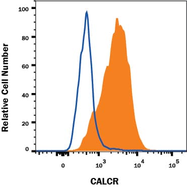
Detection of Calcitonin R in MCF‑7 Human Cell Line by Flow Cytometry.
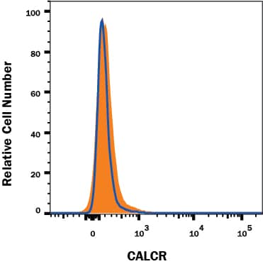
MCF-7 human breast cancer cell line was stained with Mouse Anti-Human Calcitonin R Monoclonal Antibody (Catalog # MAB4614, filled histogram) or isotype control antibody (Catalog # MAB003, open histogram), followed by Phycoerythrin-conjugated Anti-Mouse IgG F(ab')2Secondary Antibody (Catalog # F0102B).Calcitonin R Specificity is Shown by Flow Cytometry in Knockout Cell Line.
Calcitonin R knockout MCF-7 human breast cancer cell line was stained with Mouse Anti-Human Calcitonin R Monoclonal Antibody (Catalog # MAB4614, filled histogram) or isotype control antibody (Catalog # MAB003, open histogram) followed by anti-Mouse IgG PE-conjugated secondary antibody (Catalog # F0102B). No staining in the Calcitonin R knockout MCF-7 cell line was observed. View our protocol for Staining Membrane-associated Proteins.Detection of Human Calcitonin R by Immunocytochemistry/Immunofluorescence
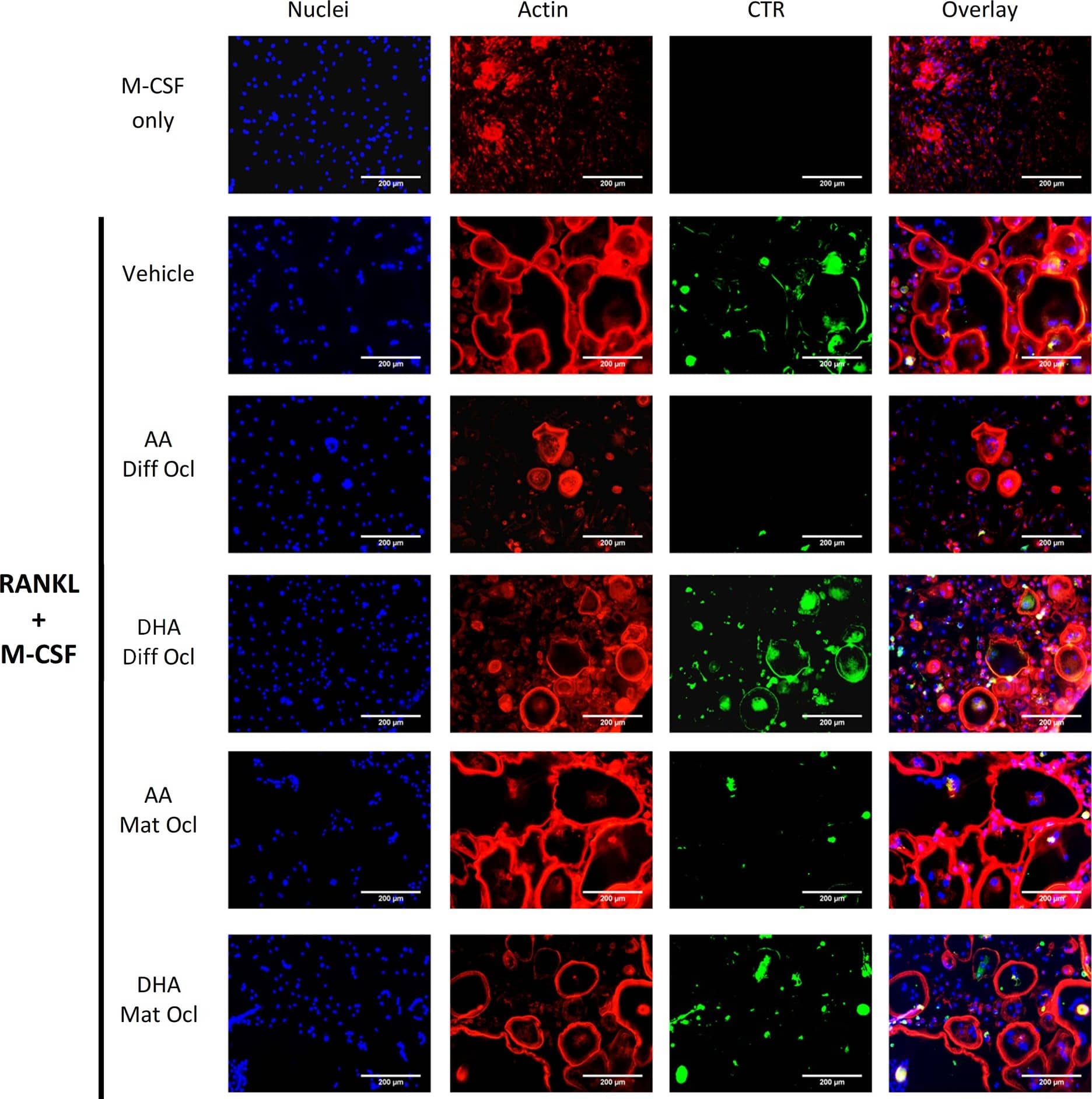
Effects of AA and DHA on actin ring formation and CTR expression.Osteoclast differentiation was stimulated in CD14+ monocytes by the addition of 25 ng ml-1 M-CSF and 30 ng ml-1 RANKL as described in Materials and Methods. Vehicle (0.08% ethanol) or LCPUFAs (40 μM) were added from day 3 in differentiating osteoclasts and from the onset of resorption (day 12–14) in mature osteoclasts. After 3 weeks of culture, cells were fixed and stained with Hoechst (blue) for nuclei, phalloidin (red) for actin rings, and anti-CTR antibody (green). The results are representative of three independent experiments conducted in triplicate. Scale bar = 200 μm. Diff Ocl—differentiating osteoclasts. Mat Ocl—mature osteoclasts. Image collected and cropped by CiteAb from the following publication (https://pubmed.ncbi.nlm.nih.gov/25867515), licensed under a CC-BY license. Not internally tested by R&D Systems.Applications for Human Calcitonin R Antibody
CyTOF-ready
Flow Cytometry
Sample: MCF-7 human breast cancer cell line
Knockout Validated
Formulation, Preparation, and Storage
Purification
Reconstitution
Formulation
Shipping
Stability & Storage
- 12 months from date of receipt, -20 to -70 °C as supplied.
- 1 month, 2 to 8 °C under sterile conditions after reconstitution.
- 6 months, -20 to -70 °C under sterile conditions after reconstitution.
Background: Calcitonin R
Calcitonin receptor (CALCR) is a glycosylated 70 kDa seven-transmembrane G protein-coupled receptor that mediates the hypocalcemic effects of the peptide hormone, calcitonin. CALCR activation inhibits osteoclast-mediated bone resorption and enhances renal calcium excretion. CALCR polymorphisms and mutations have been associated with several bone disorders. Alternative splicing results in the deletion of 16 aa in the first cytoplasmic loop or 23 aa in the first extracellular region. Human CALCR shares 70%‑72% aa sequence identity with mouse and rat CACLR.
Long Name
Alternate Names
Gene Symbol
UniProt
Additional Calcitonin R Products
Product Documents for Human Calcitonin R Antibody
Product Specific Notices for Human Calcitonin R Antibody
For research use only

