Human CD4 Antibody
R&D Systems, part of Bio-Techne | Catalog # MAB379


Key Product Details
Species Reactivity
Validated:
Cited:
Applications
Validated:
Cited:
Label
Antibody Source
Product Specifications
Immunogen
Extracellular domain
Specificity
Clonality
Host
Isotype
Scientific Data Images for Human CD4 Antibody
CD4 in Human PBMCs.
CD4 was detected in immersion fixed human peripheral blood mononuclear cells (PBMCs) using Mouse Anti-Human CD4 Monoclonal Antibody (Catalog # MAB379) at 10 µg/mL for 3 hours at room temperature. Cells were stained using the NorthernLights™ 493-conjugated Anti-Mouse IgG Secondary Antibody (green; Catalog # NL009) and counterstained with DAPI(blue). Specific staining was localized to the cell surface. View our protocol for Fluorescent ICC Staining of Non-adherent Cells.Detection of Human CD4 by Immunocytochemistry/Immunofluorescence
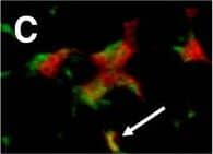
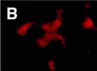
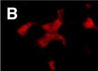
CX3CR1 expressions in affected muscle of patients with polymyositis. The muscle tissues of polymyositis (PM) patients were double-stained with CD4, CD8, or CD68, as well as CX3CR1, and were analyzed with fluorescent microscopy: (A) CX3CR1, (B) CD4, (C) merged (A) and (B), (D) CX3CR1, (E) CD8, (F) merged (D) and (E), (G) CX3CR1, (H) CD68, and (I) merged (G) and (H). Arrows indicate double-positive cells. Original magnification, ×400. Image collected and cropped by CiteAb from the following publication (https://arthritis-research.biomedcentral.com/articles/10.1186/ar3761), licensed under a CC-BY license. Not internally tested by R&D Systems.Detection of Human CD4 by Immunocytochemistry/Immunofluorescence
CX3CR1 expressions in affected muscle of patients with polymyositis. The muscle tissues of polymyositis (PM) patients were double-stained with CD4, CD8, or CD68, as well as CX3CR1, and were analyzed with fluorescent microscopy: (A) CX3CR1, (B) CD4, (C) merged (A) and (B), (D) CX3CR1, (E) CD8, (F) merged (D) and (E), (G) CX3CR1, (H) CD68, and (I) merged (G) and (H). Arrows indicate double-positive cells. Original magnification, ×400. Image collected and cropped by CiteAb from the following publication (https://arthritis-research.biomedcentral.com/articles/10.1186/ar3761), licensed under a CC-BY license. Not internally tested by R&D Systems.Applications for Human CD4 Antibody
CyTOF-ready
Flow Cytometry
Sample: Human whole blood lymphocytes
Immunocytochemistry
Sample: Immersion fixed human peripheral blood mononuclear cells
Reviewed Applications
Read 2 reviews rated 4 using MAB379 in the following applications:
Formulation, Preparation, and Storage
Purification
Reconstitution
Formulation
Shipping
Stability & Storage
- 12 months from date of receipt, -20 to -70 °C as supplied.
- 1 month, 2 to 8 °C under sterile conditions after reconstitution.
- 6 months, -20 to -70 °C under sterile conditions after reconstitution.
Background: CD4
CD4 is an approximately 55 kDa type I membrane glycoprotein that is expressed predominantly on most thymocytes and a subset of mature T lymphocytes. In humans, CD4 is also expressed to a lesser extent on monocytes and macrophage related cells. Human CD4 cDNA encodes a 458 amino acid (aa) precursor protein with a 25 aa signal peptide, a 371 aa extracellular region containing four immunoglobulin homology domains, a 24 aa transmembrane domain and a 38 aa cytoplasmic domain. CD4 is a coreceptor required for T cell recognition of antigens that are presented by class II major histocompatibility complexes. CD4 has been shown to be a coreceptor of HIV entry and specifically binds gp120, the external envelope glycoprotein of HIV.
References
- Capon, D.I. et al. (1991) Annu. Rev. Immunol. 9:649.
Alternate Names
Entrez Gene IDs
Gene Symbol
Additional CD4 Products
Product Documents for Human CD4 Antibody
Product Specific Notices for Human CD4 Antibody
For research use only


