Human Cystatin E/M Antibody
R&D Systems, part of Bio-Techne | Catalog # MAB1286

Key Product Details
Species Reactivity
Validated:
Cited:
Applications
Validated:
Cited:
Label
Antibody Source
Product Specifications
Immunogen
Arg29-Met149
Accession # Q15828
Specificity
Clonality
Host
Isotype
Scientific Data Images for Human Cystatin E/M Antibody
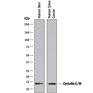
Detection of Human Cystatin E/M by Western Blot.
Western blot shows lysates of human skin tissue and human colon cancer tissue. PVDF membrane was probed with 2 µg/mL of Mouse Anti-Human Cystatin E/M Monoclonal Antibody (Catalog # MAB1286) followed by HRP-conjugated Anti-Mouse IgG Secondary Antibody (Catalog # HAF007). A specific band was detected for Cystatin E/M at approximately 13 kDa (as indicated). This experiment was conducted under reducing conditions and using Immunoblot Buffer Group 1.Cystatin E/M in Human Skin.
Cystatin E/M was detected in immersion fixed paraffin-embedded sections of human skin using 25 µg/mL Mouse Anti-Human Cystatin E/M Monoclonal Antibody (Catalog # MAB1286) overnight at 4 °C. Tissue was stained with the Anti-Mouse HRP-DAB Cell & Tissue Staining Kit (brown; Catalog # CTS002) and counterstained with hematoxylin (blue). Specific labeling was localized to the cytoplasm of cells in hair follicules. View our protocol for Chromogenic IHC Staining of Paraffin-embedded Tissue Sections.Applications for Human Cystatin E/M Antibody
Immunohistochemistry
Sample: Immersion fixed paraffin-embedded sections of human skin
Immunoprecipitation
Sample: Conditioned cell culture medium spiked with Recombinant Human Cystatin E/M (Catalog # 1286-PI), see our available Western blot detection antibodies
Western Blot
Sample: Human skin tissue and human colon cancer tissue
Formulation, Preparation, and Storage
Purification
Reconstitution
Formulation
Shipping
Stability & Storage
- 12 months from date of receipt, -20 to -70 °C as supplied.
- 1 month, 2 to 8 °C under sterile conditions after reconstitution.
- 6 months, -20 to -70 °C under sterile conditions after reconstitution.
Background: Cystatin E/M
Cystatin E/M encoded by the CST6 gene is a member of family 2 of the cystatin superfamily (1, 2). It inhibits papain and cathepsin B, two of the cysteine proteases. Its mRNA was found in many tissues by the two groups who did initial cloning (1, 2). However, its protein was found only in skin and sweat glands by a third group (3). In addition to being a cysteine protease inhibitor, Cystatin E/M is also a substrate for transglutaminases (3). It is required for viability and for correct formation of cornified layers in the epidermis and hair follicles, as ichq mice, with a null mutation in the Cystatin E/M gene, have defects in epidermal cornification and die between 5 and 12 days of age (4). Cystatin E/M expression and function may not be limited to cutaneous epithelia. For example, it is found in rat brain and is induced during neuronal cell differentiation (5).
References
- Sotiropoulou, G. et al. (1997) J. Biol. Chem. 272:903.
- Ni, J. et al. (1997) J. Biol. Chem. 272:10853.
- Zeeuwen, P.L. et al. (2001) J. Invest. Dermatol. 116:693.
- Zeeuwen, P.L. et al. (2002) Hum. Mol. Genet. 11:2867.
- Hong, J. et al. (2002) J. Neurochem. 81:922.
Alternate Names
Gene Symbol
UniProt
Additional Cystatin E/M Products
Product Documents for Human Cystatin E/M Antibody
Product Specific Notices for Human Cystatin E/M Antibody
For research use only

