Human eNOS Antibody
R&D Systems, part of Bio-Techne | Catalog # AF950


Key Product Details
Species Reactivity
Validated:
Cited:
Applications
Validated:
Cited:
Label
Antibody Source
Product Specifications
Immunogen
Specificity
Clonality
Host
Isotype
Scientific Data Images for Human eNOS Antibody
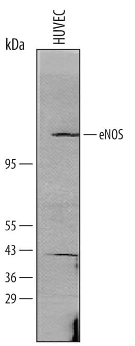
Detection of Human eNOS by Western Blot.
Western blot shows lysates of HUVEC human umbilical vein endothlial cells. PVDF membrane was probed with 1 µg/mL of Goat Anti-Human eNOS Affinity-purified Polyclonal Antibody (Catalog # AF950) followed by HRP-conjugated Anti-Goat IgG Secondary Antibody (Catalog # HAF109). A specific band was detected for eNOS at approximately 130-140 kDa (as indicated). This experiment was conducted under reducing conditions and using Immunoblot Buffer Group 8.Immunoprecipitation of Human eNOS.
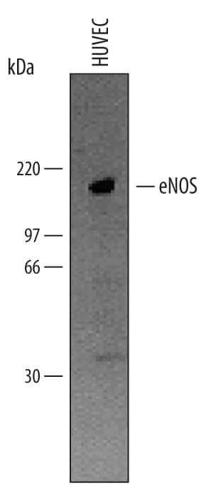
eNOS was immunoprecipitated from lysates (1 x 10⁶cells) of HUVEC human umbilical vein endothelial cells following incubation with 0.3 µg Goat Anti-Human eNOS Antigen Affinity-purified Polyclonal Antibody (Catalog # AF950). Immunoprecipitated eNOS was detected by Western blot using 1 µg/mL Human eNOS Antigen Affinity-purified Polyclonal Antibody (Catalog # AF950). View our recommended buffer recipes for immunoprecipitation.Detection of Human eNOS by Simple WesternTM.
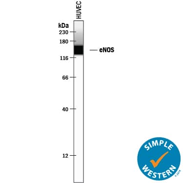
Simple Western lane view shows lysates of HUVEC human umbilical vein endothelial cells, loaded at 0.2 mg/mL. A specific band was detected for eNOS at approximately 139 kDa (as indicated) using 10 µg/mL of Goat Anti-Human eNOS Antigen Affinity-purified Polyclonal Antibody (Catalog # AF950) followed by 1:50 dilution of HRP-conjugated Anti-Goat IgG Secondary Antibody (Catalog # HAF109). This experiment was conducted under reducing conditions and using the 12-230 kDa separation system.Applications for Human eNOS Antibody
Immunohistochemistry
Sample: Immersion fixed paraffin-embedded sections of human placenta subjected to Antigen Retrieval Reagent-Basic (Catalog # CTS013)
Immunoprecipitation
Sample: HUVEC human umbilical vein endothelial cells, see our available Western blot detection antibodies
Simple Western
Sample: HUVEC human umbilical vein endothelial cells
Western Blot
Sample: eNOS immunoprecipitated HUVEC human umbilical vein endothelial cells
Formulation, Preparation, and Storage
Purification
Reconstitution
Formulation
Shipping
Stability & Storage
- 12 months from date of receipt, -20 to -70 °C as supplied.
- 1 month, 2 to 8 °C under sterile conditions after reconstitution.
- 6 months, -20 to -70 °C under sterile conditions after reconstitution.
Background: eNOS
Endothelial NOS (eNOS), also known as nitric oxide synthase 3 (NOS3) or constitutive NOS (cNOS), is an enzyme encoded by the NOS3 gene. Endothelial NOS generates nitric oxide in blood vessels and is involved with regulating vascular tonehttp://en.wikipedia.org/wiki/Vascular_resistance" title="Vascular resistance"> by inhibiting smooth muscle contraction and platelethttp://en.wikipedia.org/wiki/Platelet" title="Platelet"> aggregation. A constitutive calciumhttp://en.wikipedia.org/wiki/Calcium_in_biology" title="Calcium in biology"> dependent NOS provides a basal release of NO. eNOS is associated with plasma membranes surrounding cells and the membranes of the Golgi apparatushttp://en.wikipedia.org/wiki/Golgi_apparatus" title="Golgi apparatus"> within cells.
Long Name
Alternate Names
Gene Symbol
Additional eNOS Products
Product Documents for Human eNOS Antibody
Product Specific Notices for Human eNOS Antibody
For research use only

