Human ESAM APC-conjugated Antibody
R&D Systems, part of Bio-Techne | Catalog # FAB4204A


Key Product Details
Species Reactivity
Applications
Label
Antibody Source
Product Specifications
Immunogen
Gln30-Ala247
Accession # Q96AP7
Specificity
Clonality
Host
Isotype
Scientific Data Images for Human ESAM APC-conjugated Antibody
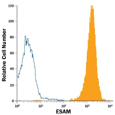
Detection of ESAM in HUVEC Human Cell Line by Flow Cytometry.
HUVEC human umbilical vein endothelial cell line was stained with Mouse Anti-Human ESAM APC-conjugated Monoclonal Antibody (Catalog # FAB4204A, filled histogram) or isotype control antibody (Catalog # IC0041A, open histogram). View our protocol for Staining Membrane-associated Proteins.Applications for Human ESAM APC-conjugated Antibody
Flow Cytometry
Sample: HUVEC human umbilical vein endothelial cell line
Formulation, Preparation, and Storage
Purification
Formulation
Shipping
Stability & Storage
Background: ESAM
Endothelial cell-selective adhesion molecule (ESAM) is a 55 kDa type I transmembrane glycoprotein that belongs to the JAM family of immunoglobulin superfamily molecules (1, 2). Human ESAM is synthesized as a 390 amino acid (aa) protein composed of a 29 aa signal peptide, a 216 aa extracellular region, a putative 26 aa transmembrane segment, and a 119 aa cytoplasmic domain. The extracellular region contains one V-type and one C2-type Ig domain and is involved in homophilic adhesion (1). In the cytoplasmic domain, there is a docking site for the multifunctional adaptor protein MAGI-1 (3). The extracellular region of human ESAM shows 90%, 74%, 69%, and 67% aa identity with monkey, canine, mouse, and rat extracellular ESAM, respectively. ESAM is expressed on endothelial cells, activated platelets, and megakaryocytes and can be found associated with cell-to-cell junctions. Whether ESAM is restricted to a particular junctional type is not clear (1, 2). ESAM deficient mice have no defect in vascularization but do have reduced angiogenic potential. This may be due to a decreased migratory response to FGF-2 (4).
References
- Hirata, K-I. et al. (2001) J. Biol. Chem. 276:16223.
- Nasdala, I. et al. (2002) J. Biol. Chem. 277:16294.
- Wegmann, F. et al. (2004) Exp. Cell Res. 300:121.
- Ishida, T. et. al. (2003) J. Biol. Chem. 278:34598.
Long Name
Alternate Names
Gene Symbol
UniProt
Additional ESAM Products
Product Documents for Human ESAM APC-conjugated Antibody
Product Specific Notices for Human ESAM APC-conjugated Antibody
For research use only