Human FGFR1 (IIIb) Antibody
R&D Systems, part of Bio-Techne | Catalog # MAB765

Key Product Details
Validated by
Biological Validation
Species Reactivity
Validated:
Human
Cited:
Human
Applications
Validated:
Neutralization, Western Blot
Cited:
Bioassay, Neutralization
Label
Unconjugated
Antibody Source
Monoclonal Mouse IgG1 Clone # 133111
Product Specifications
Immunogen
A mixture of purified isoforms of recombinant human FGF R1 extracellular domains
Specificity
Detects IIIb isoforms of human FGF R1. In direct ELISAs and Western blots, this antibody detected IIIb but not IIIc isoforms of human FGF R1, human FGF R2, and mouse FGF R2. In direct ELISA, approximately 20% cross-reactivity was observed with recombinant human (rh) FGF R3 (IIIb). No cross-reactivity was observed with rhFGF R4 or with IIIIc isoforms of rhFGF R1, rhFGF R2, rmFGF R2, or rhFGF R3.
Clonality
Monoclonal
Host
Mouse
Isotype
IgG1
Endotoxin Level
<0.10 EU per 1 μg of the antibody by the LAL method.
Scientific Data Images for Human FGFR1 (IIIb) Antibody
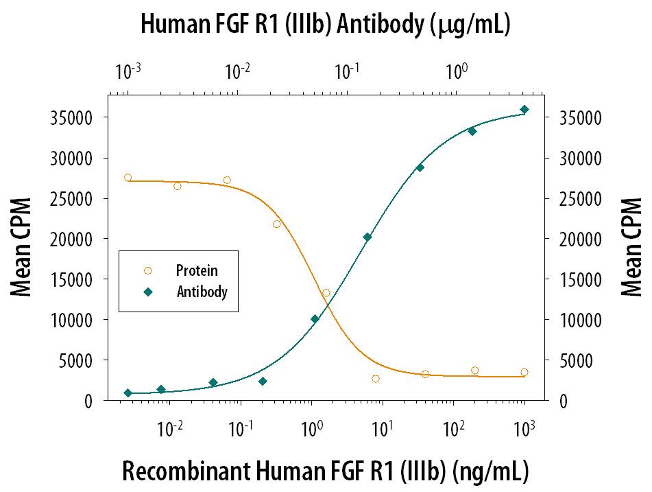
FGF R1 alpha Inhibition of FGF acidic-dependent Cell Proliferation and Neutralization by Human FGF R1 Antibody.
Recombinant Human FGF R1a (IIIb) Fc Chimera (Catalog # 655-FR) inhibits Recombinant Human FGF acidic (Catalog # 232-FA) induced proliferation in the NR6R-3T3 mouse fibroblast cell line in a dose-dependent manner (orange line). Inhibition of Recombinant Human FGF acidic (0.3 ng/mL) activity elicited by Recombinant Human FGF R1a (IIIb) Fc Chimera (6 ng/mL) is neutralized (green line) by increasing concentrations of Human FGF R1 (IIIb) Monoclonal Antibody (Catalog # MAB765). The ND50 is typically 0.075-0.3 µg/mL in the presence of heparin (10 µg/mL).Detection of Human FGFR1 by Immunocytochemistry/Immunofluorescence
Proximity between AcSDKP and FGFR1 inhibits the TGF beta/smad signaling pathway in HMVECs. (a) HMVECs were treated with N-FGFR1 (1.5 μg/ml) for 48 h with or without preincubation with AcSDKP (100 nM) for 2 h, and the proximity between AcSDKP and FGFR1 was analyzed by the Duolink In Situ Assay. For each slide, images at a × 400 original magnification were obtained from six different areas. (b and c) HMVECs were treated with TGF beta2 (5 ng/ml) for 15 min or 48 h with or without preincubation with AcSDKP for 2 h, and the p-smad3, TGF betaR1, TGF betaR2 and FGFR1 levels were analyzed by western blot. Densitometric analysis of the p-smad3/smad3, TGF betaR1/ beta-actin, TGF betaR2/ beta-actin and FGFR1/ beta-actin levels from each group (n=6) were analyzed. (d and e) HMVECs were incubated with TGF beta2 for 15 min or 48 h with or without preincubation with AcSDKP or its mutants (AcDSPK, AcSDKA, AcADKP) (100 nM) for 2 h. The p-smad3/smad3, TGF betaR1/ beta-actin, TGF betaR2/ beta-actin and FGFR1/ beta-actin protein levels were analyzed by western blot Image collected and cropped by CiteAb from the following publication (https://pubmed.ncbi.nlm.nih.gov/28771231), licensed under a CC-BY license. Not internally tested by R&D Systems.Detection of Human FGFR1 by Western Blot
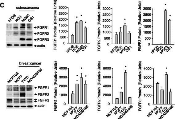
Expression of FGFR1 in cells from breast and bone tissue. a, FGFR mRNA transcript levels in normal and cancer cells. b, Fold change in FGFR mRNA based on expression in cancers cells relative to normal cells. c, FGFR protein levels in normal versus cancer cells. Results are from triplicate experiments and the Western blot is representative of the triplicates. (*p < 0.05) Image collected and cropped by CiteAb from the following open publication (https://pubmed.ncbi.nlm.nih.gov/26201468), licensed under a CC-BY license. Not internally tested by R&D Systems.Applications for Human FGFR1 (IIIb) Antibody
Application
Recommended Usage
Western Blot
1 µg/mL
Sample: Recombinant Human FGF R1 alpha (IIIb) Fc Chimera (Catalog # 655-FR)
Sample: Recombinant Human FGF R1 alpha (IIIb) Fc Chimera (Catalog # 655-FR)
Neutralization
Measured by its ability to neutralize FGF R1 alpha-mediated inhibition of proliferation in the NR6R-3T3 mouse fibroblast cell line. The Neutralization Dose (ND50) is typically 0.075-0.3 µg/mL in the presence of 6 ng/mL Recombinant Human FGF R1 alpha (IIIb) Fc Chimera, 0.3 ng/mL Recombinant Human FGF acidic, and 10 µg/mL heparin.
Formulation, Preparation, and Storage
Purification
Protein A or G purified from hybridoma culture supernatant
Reconstitution
Reconstitute at 0.5 mg/mL in sterile PBS. For liquid material, refer to CoA for concentration.
Formulation
Lyophilized from a 0.2 μm filtered solution in PBS with Trehalose. *Small pack size (SP) is supplied either lyophilized or as a 0.2 µm filtered solution in PBS.
Shipping
Lyophilized product is shipped at ambient temperature. Liquid small pack size (-SP) is shipped with polar packs. Upon receipt, store immediately at the temperature recommended below.
Stability & Storage
Use a manual defrost freezer and avoid repeated freeze-thaw cycles.
- 12 months from date of receipt, -20 to -70 °C as supplied.
- 1 month, 2 to 8 °C under sterile conditions after reconstitution.
- 6 months, -20 to -70 °C under sterile conditions after reconstitution.
Background: FGFR1
Long Name
Fibroblast Growth Factor Receptor 1
Alternate Names
CD331, FGF R1, Flt-2
Gene Symbol
FGFR1
Additional FGFR1 Products
Product Documents for Human FGFR1 (IIIb) Antibody
Product Specific Notices for Human FGFR1 (IIIb) Antibody
For research use only
Loading...
Loading...
Loading...
Loading...
Loading...


