Human GPR115 Antibody
R&D Systems, part of Bio-Techne | Catalog # MAB54371


Key Product Details
Species Reactivity
Applications
Label
Antibody Source
Product Specifications
Immunogen
Ser22-Ala347
Accession # Q8IZF3
Specificity
Clonality
Host
Isotype
Scientific Data Images for Human GPR115 Antibody
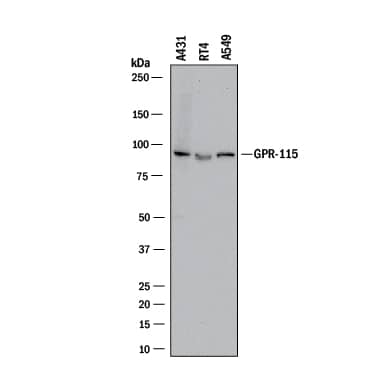
Detection of Human GPR115 by Western Blot.
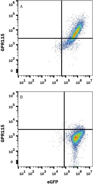
Western blot shows lysates of A431 human epithelial carcinoma cell line, RT-4 human bladder carcinoma cell line, and A549 human lung carcinoma cell line. PVDF membrane was probed with 2 µg/mL of Mouse Anti-Human GPR115 Monoclonal Antibody (Catalog # MAB54371) followed by HRP-conjugated Anti-Mouse IgG Secondary Antibody (Catalog # HAF018). A specific band was detected for GPR115 at approximately 90 kDa (as indicated). This experiment was conducted under reducing conditions and using Immunoblot Buffer Group 1.Detection of GPR115 in HEK293 Human Cell Line Transfected with Human GPR115 and eGFP by Flow Cytometry
HEK293 human embryonic kidney cell line transfected with (A) human GPR115 or (B) irrelevant protein and eGFP was stained with Mouse Anti-Human GPR115 Monoclonal Antibody (Catalog # MAB54371) followed by APC-conjugated Anti-Mouse IgG Secondary Antibody (Catalog # F0101B). Quadrant markers were set based on control antibody staining (Catalog # MAB002). View our protocol for Staining Membrane-associated Proteins.Applications for Human GPR115 Antibody
CyTOF-ready
Flow Cytometry
Sample: HEK293 Human Cell Line Transfected with Human GPR115 and eGFP
Western Blot
Sample: A431 human epithelial carcinoma cell line, RT-4 human bladder carcinoma cell line, and A549 human lung carcinoma cell line
Formulation, Preparation, and Storage
Purification
Reconstitution
Formulation
Shipping
Stability & Storage
- 12 months from date of receipt, -20 to -70 °C as supplied.
- 1 month, 2 to 8 °C under sterile conditions after reconstitution.
- 6 months, -20 to -70 °C under sterile conditions after reconstitution.
Background: GPR115
GPR115 is a member of the LN-7TM family of adhesion-type 7-transmembrane (TM) G-protein coupled receptors (GPCR) that show a long extracellular N-terminus (1, 2). The 695 amino acid (aa) human GPR115 sequence predicts a 21 aa signal sequence, a 385 aa N‑terminal extracellular domain (ECD), seven TM regions separated by 6‑24 aa intracellular and extracellular regions, and a 40 aa cytoplasmic tail. Like other LN-7TM members, the ECD contains a highly glycosylated mucin‑like stalk that is predicted to function in adhesion. This is followed by a cysteine-rich GPCR proteolytic cleavage site (GPS) (1). GPS domains, which have been described in other 7TM proteins including ETL, GPR126, HE6, and Latrophilin-1, are cleavage sites for processing proteins into two subunits (3‑7). Within the N‑terminal region that ends with the predicted cleavage site (aa 22‑347), human GPR115 shares 58% aa sequence identity with the corresponding region of mouse and rat GPR115. GPR115 was identified from expressed sequence tags (ESTs) found in pregnant uterus, breast, and the genitourinary tract (1).
References
- Fredriksson, R. et al. (2002) FEBS Lett. 531:407.
- Bjarnadottir, T.K. et al. (2004) Genomics 84:23.
- Nechiporuk, T. et al. (2001) J. Biol. Chem. 276:4150.
- Moriguchi, T. et al. (2004) Genes Cells 9:549.
- Kierszenbaum, A.L. (2003) Mol. Reprod. Dev. 64:1.
- Krasnoperov, V.G. et al. (1997) Neuron 18:925.
- Krasnoperov, V. et al. (2002) J. Biol. Chem. 277:46518.
Long Name
Alternate Names
Gene Symbol
UniProt
Additional GPR115 Products
Product Documents for Human GPR115 Antibody
Product Specific Notices for Human GPR115 Antibody
For research use only
