Human IL-31 Antibody
R&D Systems, part of Bio-Techne | Catalog # AF2824


Key Product Details
Validated by
Species Reactivity
Validated:
Cited:
Applications
Validated:
Cited:
Label
Antibody Source
Product Specifications
Immunogen
Ser24-Thr164
Accession # Q6EBC2
Specificity
Clonality
Host
Isotype
Endotoxin Level
Scientific Data Images for Human IL-31 Antibody
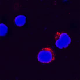
IL‑31 in Human PBMCs.
IL-31 was detected in immersion fixed human peripheral blood mononuclear cells (PBMCs) treated with calcium ionomycin and PMA using Goat Anti-Human IL-31 Antigen Affinity-purified Polyclonal Antibody (Catalog # AF2824) at 15 µg/mL for 3 hours at room temperature. Cells were stained using the NorthernLights™ 557-conjugated Anti-Goat IgG Secondary Antibody (red; Catalog # NL001) and counterstained with DAPI (blue). Specific staining was localized to cell surfaces and cytoplasm. View our protocol for Fluorescent ICC Staining of Non-adherent Cells.Applications for Human IL-31 Antibody
Immunocytochemistry
Sample: Immersion fixed human peripheral blood mononuclear cells treated with calcium ionomycin and PMA
Western Blot
Sample: Recombinant Human IL-31 (Catalog # 2824-IL)
Neutralization
Human IL-31 Sandwich Immunoassay
Formulation, Preparation, and Storage
Purification
Reconstitution
Formulation
Shipping
Stability & Storage
- 12 months from date of receipt, -20 to -70 °C as supplied.
- 1 month, 2 to 8 °C under sterile conditions after reconstitution.
- 6 months, -20 to -70 °C under sterile conditions after reconstitution.
Background: IL-31
Human Interleukin-31 (IL-31) is a 24 kDa, short-chain member of the alpha-helical family of cytokines. The human IL-31 cDNA encodes a 164 amino acid (aa) precursor that contains a 23 aa signal peptide and a 141 aa mature protein (1, 2). The mature region shows four alpha-helices which would be expected to show a typical up‑up‑down‑down topology. Human and mouse IL-31 share 24% aa sequence identity in the mature region (1). IL-31 is mainly associated with activated T cells and preferentially expressed by Th2 rather than Th1 cells. IL-31 signals via a heterodimeric receptor complex composed of a 120 kDa, gp130-related molecule termed IL‑31 RA (also GPL and GLM-R) and the 180 kDa oncostatin M receptor (OSM R beta) (2‑6). In the complex, IL-31 directly binds to GPL, not OSM R (2, 3). IL-31 signaling has been shown to involve the Jak/STAT pathway, the PI3 kinase/AKT cascade, and the MAP kinase pathway (2‑5). Although multiple isoforms of IL-31 RA are known, only a form that contains the entire length of the cytoplasmic domain is signaling-capable (2, 3). The IL-31 receptor is constitutively expressed by keratinocytes and upregulated by IFN-gamma on monocytes (1). Studies using transgenic mice indicate that IL-31 may contribute to the pruritis (itching) associated with nonatopic dermatitis (1, 7).
References
- Dillon, S.R. et al. (2004) Nat. Immunol. 5:752.
- Diveu, C. et al. (2004) Eur. Cytokine Netw. 15:291.
- Dreuw, A. et al. (2004) J. Biol. Chem. 279:36112.
- Diveu, C. et al. (2003) J. Biol. Chem. 278:49850.
- Ghilardi, N. et al. (2002) J. Biol. Chem 277:16831.
- Mosley, B. et al. (1996) J. Biol. Chem. 271:32635.
- Takaoka, A. et al. (2005) Eur. J. Pharmacol. 516:180.
Long Name
Alternate Names
Gene Symbol
UniProt
Additional IL-31 Products
Product Documents for Human IL-31 Antibody
Product Specific Notices for Human IL-31 Antibody
For research use only