Human IL-4I1 Antibody
R&D Systems, part of Bio-Techne | Catalog # MAB5684

Key Product Details
Validated by
Species Reactivity
Applications
Label
Antibody Source
Product Specifications
Immunogen
Met1-His567
Accession # Q96RQ9
Specificity
Clonality
Host
Isotype
Scientific Data Images for Human IL-4I1 Antibody
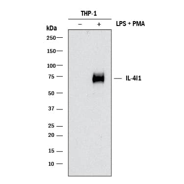
Detection of Human IL‑4I1 by Western Blot.
Western blot shows lysates of THP-1 human acute monocytic leukemia cell line untreated (-) or treated (+) with 200 nM PMA for 24 hours and 10 µg/mL LPS for 3 hours. PVDF membrane was probed with 2 µg/mL of Rat Anti-Human IL-4I1 Monoclonal Antibody (Catalog # MAB5684) followed by HRP-conjugated Anti-Rat IgG Secondary Antibody (Catalog # HAF005). A specific band was detected for IL-4I1 at approximately 75 kDa (as indicated). This experiment was conducted under reducing conditions and using Immunoblot Buffer Group 1.IL‑4I1 in HDLM‑2 Human Cell Line.
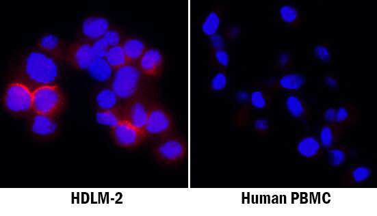
IL-4I1 was detected in immersion fixed HDLM-2 human Hodgkin's lymphoma cell line using Rat Anti-Human IL-4I1 Monoclonal Antibody (Catalog # MAB5684) at 8 µg/mL for 3 hours at room temperature. Cells were stained using the NorthernLights™ 557-conjugated Anti-Rat IgG Secondary Antibody (red; Catalog # NL013) and counterstained with DAPI (blue). Specific staining was localized to cytoplasm in lysosomes. View our protocol for Fluorescent ICC Staining of Non-adherent Cells.IL‑4I1 in Human B Cell Lymphoma.
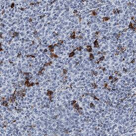
IL-4I1 was detected in immersion fixed paraffin-embedded sections of human B cell lymphoma using Rat Anti-Human IL-4I1 Monoclonal Antibody (Catalog # MAB5684) at 5 µg/mL for 1 hour at room temperature followed by incubation with the Anti-Rat IgG VisUCyte™ HRP Polymer Antibody (Catalog # VC005). Before incubation with the primary antibody, tissue was subjected to heat-induced epitope retrieval using Antigen Retrieval Reagent-Basic (Catalog # CTS013). Tissue was stained using DAB (brown) and counterstained with hematoxylin (blue). Specific staining was localized to cytoplasm in lymphocytes. View our protocol for IHC Staining with VisUCyte HRP Polymer Detection Reagents.Applications for Human IL-4I1 Antibody
Immunocytochemistry
Sample: Immersion fixed HDLM-2 human Hodgkin's lymphoma cell line
Immunohistochemistry
Sample: Immersion fixed paraffin-embedded sections of human B cell lymphoma
Western Blot
Sample: THP‑1 human acute monocytic leukemia cell line treated in PMA and LPS
Reviewed Applications
Read 1 review rated 5 using MAB5684 in the following applications:
Formulation, Preparation, and Storage
Purification
Reconstitution
Formulation
Shipping
Stability & Storage
- 12 months from date of receipt, -20 to -70 °C as supplied.
- 1 month, 2 to 8 °C under sterile conditions after reconstitution.
- 6 months, -20 to -70 °C under sterile conditions after reconstitution.
Background: IL-4I1
Interleukin 4 induced protein 1 (IL-4I1), also known as protein FIG-1 and L-amino acid oxidase, is encoded by a B-cell IL-4-inducible gene, FIG1, and is highly expressed in primary metastinal B-cell lymphomas (1-4). It belongs to the flavin monoamine oxidase family, FIG1 subfamily. Enzymological characterization reveals that IL-4I1 has L-amino acid oxidase activity with preference toward aromatic amino acids. Studies have shown that hIL-4I1 inhibited the proliferation of CD3‑stimulated T lymphocytes with a similar effect on CD4(+) and CD8(+) T cells (5). Its inhibitory effect was dependent on enzymatic activity and H2O2 production. Its restricted expression to lymphoid tissues indicates that it may play an important function in the immune system (1, 4).
References
- Chu, C.C. and W.E. Paul. (1997) Proc. Natl. Acad. Sci. USA 94:2507.
- Mason, J.M. et al. (2004) J. Immunol. 173:4561.
- Chavan, S.S. et al. (2002) Biochim. Biophys. Acta. 1576:70.
- Copie-Bergman, C. et al. (2003) Blood 101:2756.
- Boulland, M.L. et al. (2007) Blood 110:220.
Long Name
Alternate Names
Gene Symbol
UniProt
Additional IL-4I1 Products
Product Documents for Human IL-4I1 Antibody
Product Specific Notices for Human IL-4I1 Antibody
For research use only


