Human LC3A Antibody
R&D Systems, part of Bio-Techne | Catalog # MAB8558

Key Product Details
Validated by
Species Reactivity
Applications
Label
Antibody Source
Product Specifications
Immunogen
Accession # Q9H492
Specificity
Clonality
Host
Isotype
Scientific Data Images for Human LC3A Antibody
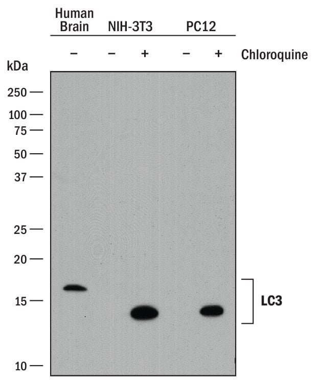
Detection of Human, Mouse, and Rat LC3A by Western Blot.
Western blot shows lysates of human brain tissue, NIH-3T3 mouse embryonic fibroblast cell line, and PC-12 rat adrenal pheochromocytoma cell line untreated (-) or treated (+) with 50 µM Chloroquine for 18 hours. PVDF membrane was probed with 2 µg/mL of Rat Anti-Human LC3A Antibody (Catalog # MAB8558) followed by HRP-conjugated Anti-Rat IgG Secondary Antibody (Catalog # HAF005). Specific bands were detected for LC3A at approximately 14 and 16 kDa (as indicated). This experiment was conducted under reducing conditions and using Immunoblot Buffer Group 1.LC3A in Human Brain Cortex Tissue.
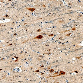
LC3A was detected in immersion fixed paraffin-embedded sections of human brain cortex tissue using Rat Anti-Human LC3A Monoclonal Antibody (Catalog # MAB8558) at 15 µg/mL overnight at 4 °C. Tissue was stained using the Anti-Rat HRP-DAB Cell & Tissue Staining Kit (brown; Catalog # CTS017) and counterstained with hematoxylin (blue). Specific staining was localized to neurons. View our protocol for Chromogenic IHC Staining of Paraffin-embedded Tissue Sections.LC3A in Human Brain.
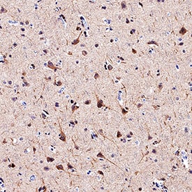
LC3A was detected in immersion fixed paraffin-embedded sections of human brain (cortex) using Rat Anti-Human LC3A Monoclonal Antibody (Catalog # MAB8558) at 1.7 µg/mL overnight at 4 °C. Tissue was stained using the Anti-Rat HRP-DAB Cell & Tissue Staining Kit (brown; Catalog # CTS017) and counterstained with hematoxylin (blue). Specific staining was localized to cytoplasm. View our protocol for Chromogenic IHC Staining of Paraffin-embedded Tissue Sections.Applications for Human LC3A Antibody
Immunohistochemistry
Sample: Immersion fixed paraffin-embedded sections of human brain cortex tissue
Western Blot
Sample: Human brain tissue (untreated), NIH‑3T3 mouse embryonic fibroblast cell line and PC‑12 rat adrenal pheochromocytoma cell line treated with Chloroquine
Reviewed Applications
Read 2 reviews rated 5 using MAB8558 in the following applications:
Formulation, Preparation, and Storage
Purification
Reconstitution
Formulation
Shipping
Stability & Storage
- 12 months from date of receipt, -20 to -70 °C as supplied.
- 1 month, 2 to 8 °C under sterile conditions after reconstitution.
- 6 months, -20 to -70 °C under sterile conditions after reconstitution.
Background: LC3A
Human Microtubule-associated Protein (MAP) Light Chain 3 (LC3) A is a121 amino acid (aa) protein with a predicted molecular weight of 14 kDa. It is a member of the LC3 subfamily of Autophagy-related 8 (Atg8) proteins (1). The LC3 subfamily also includes LC3B andLC3C. LC3 exhibits 100% aa sequence identity with its mouse and rat orthologs, and is orthologous to the yeast autophagy-related protein Atg8. Atg8 family members show structural similarity with Ubiquitin, but lack aa sequence similarity. LC3 was originally described as part is part of a complex that includes heavy and light chains comprising the MAP1 family of microtubule regulatory proteins (3). However, LC3 has gained attention for MAP1-independent functions in autophagy. LC3 utilizes a ubiquitin-like conjugation system that includes E1-, E2-, and E3-like enzymes to covalently attach phosphatidylethanolamine (PE) to its C-terminus, incorporating it into the phagophore membrane during the early stages of autophagasome formation (4). Recruitment of LC3 to the phagophore may promote membrane elongation (4,5). It may also be involved in cargo recruitment to autophagosomes (1). LC3 is often used as a marker of autophagy.
References
- Shpilka, T. et al. (2011) Genome Biol. 12:226.
- He, H. et al. (2003) J. Biol. Chem. 278:29278.
- Kuznetsov, S.A. & V.I. Gelfand (1987) FEBS Let. 212:145.
- Weidberg, H. et al. (2011) Ann Rev. Biochem. 80:125.
- Weidberg, H. et al. (2010) EMBO J. 29:1792.
Long Name
Alternate Names
Gene Symbol
UniProt
Additional LC3A Products
Product Documents for Human LC3A Antibody
Product Specific Notices for Human LC3A Antibody
For research use only


