Human MD2 Biotinylated Antibody
R&D Systems, part of Bio-Techne | Catalog # BAF1787


Key Product Details
Species Reactivity
Applications
Label
Antibody Source
Product Specifications
Immunogen
Glu17-Asn160
Accession # NP_001123946
Specificity
Clonality
Host
Isotype
Scientific Data Images for Human MD2 Biotinylated Antibody
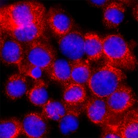
MD2 in U937 Human Cell Line.
MD2 was detected in immersion fixed U937 human histiocytic lymphoma cell line using Sheep Anti-Human MD2 Biotinylated Antigen Affinity-purified Polyclonal Antibody (Catalog # BAF1787) at 15 µg/mL for 3 hours at room temperature. Cells were stained using the NorthernLights™ 557-conjugated Streptavidin (red; NL999) and counterstained with DAPI (blue). Specific staining was localized to cytoplasm. Staining was performed using our protocol for Fluorescent ICC Staining of Non-adherent Cells.Applications for Human MD2 Biotinylated Antibody
Immunocytochemistry
Sample: Immersion fixed U937 human histiocytic lymphoma cell line
Western Blot
Sample: Recombinant Human MD-2 (Catalog # 1787-MD)
Formulation, Preparation, and Storage
Purification
Reconstitution
Formulation
Shipping
Stability & Storage
- 12 months from date of receipt, -20 to -70 °C as supplied.
- 1 month, 2 to 8 °C under sterile conditions after reconstitution.
- 6 months, -20 to -70 °C under sterile conditions after reconstitution.
Background: MD2
MD-2, also known as lymphocyte antigen 96 and ESOP-1, is a secreted glycoprotein that shares conserved cysteine residues and significant sequence similarity (23%) with MD-1. The gene of human MD-2 encodes a 160 amino acid residue (aa) precursor protein with a 16 aa signal peptide and a 144 aa mature protein, which contains 2 N-glycosylation sites (1). Recombinant secreted MD-2 has been found to exist as disulfide-linked dimers and oligomers (2).
Both MD-1 and MD-2 are accessory molecules that associate with the extracellular leucine-rich repeats (LRR) of Toll-like receptor (TLR) family members, which are type I transmembrane receptors that regulate innate immune responses to microbial pathogens (3, 4). MD-1 binds to RP105 on B cells and macrophages to form the signaling receptor complex for lipopolysaccharide (LPS), a constituent of the outer membrane of Gram-negative bacteria. Similarly, MD-2 interacts with TLR-4 to form the heteromeric receptor that confers LPS responsiveness. MD-2 also associates with TLR-2, albeit with less avidity, to confer responsiveness to cell wall components from both Gram-positive and Gram-negative bacteria. MD-1 and MD-2 are also required for the correct targeting of the TLRs to the cell surface. Although MD-2 glycosylation is not crucial for its surface expression and interaction with TLR-4, it is required for LPS binding and signaling (5).
References
- Shimazu, R. et al. (1999) J. Exp. Med. 189:1777.
- Visintin, A. et al. (2001) Proc. Natl. Acad. Sci. USA 98:12156.
- Nagai, Y. et al. (2002) Nature Immunology 3:667.
- Akashi, S. et al. (2003) J. Exp. Med. 198:1035.
- Correia, J. and R. Ulevitch (2002) J. Biol. Chem. 277:1845.
Long Name
Alternate Names
Gene Symbol
UniProt
Additional MD2 Products
Product Documents for Human MD2 Biotinylated Antibody
Product Specific Notices for Human MD2 Biotinylated Antibody
For research use only