Human/Mouse FGF-8 Antibody
R&D Systems, part of Bio-Techne | Catalog # MAB323

Key Product Details
Species Reactivity
Validated:
Human, Mouse
Cited:
Human, Mouse, Avian - Chicken
Applications
Validated:
Immunohistochemistry, Neutralization, Western Blot
Cited:
Immunocytochemistry, Immunohistochemistry, Immunohistochemistry-Paraffin, In ovo assay, Western Blot
Label
Unconjugated
Antibody Source
Monoclonal Mouse IgG1 Clone # 47109
Product Specifications
Immunogen
E. coli-derived recombinant mouse FGF-8b
Gln23-Arg215
Accession # NP_001159834
Gln23-Arg215
Accession # NP_001159834
Specificity
Detects human and mouse FGF-8 in direct ELISAs and Western blots. In direct ELISAs and Western blots, less than 2% cross-reactivity with recombinant human (rh) FGF-5, rhFGF-7, and rhFGF-9 is observed.
Clonality
Monoclonal
Host
Mouse
Isotype
IgG1
Endotoxin Level
<0.10 EU per 1 μg of the antibody by the LAL method.
Scientific Data Images for Human/Mouse FGF-8 Antibody
Cell Proliferation Induced by FGF‑8 and Neutralization by Human and Mouse FGF‑8 Antibody.
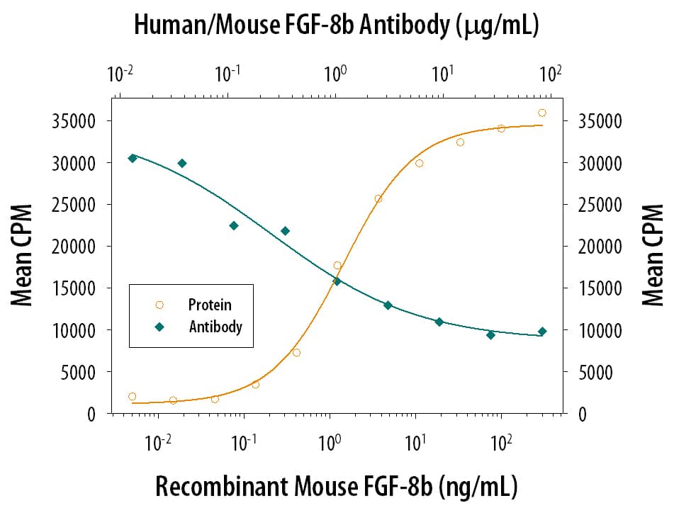
Recombinant Mouse FGF-8 c Isoform (Catalog # 424-FC) stimulates proliferation in the the NR6R-3T3 mouse fibroblast cell line in a dose-dependent manner (orange line). Proliferation elicited by Recombinant Mouse FGF-8 c Isoform (125 ng/mL) is neutralized (green line) by increasing concentrations of Mouse Anti-Human/Mouse FGF-8 Monoclonal Antibody (Catalog # MAB323). The ND50 is typically 0.25-0.75 µg/mL in the presence of heparin (0.1 µg/mL).FGF‑8 in Human Prostate.
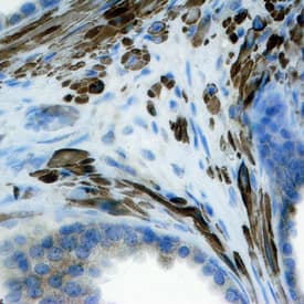
FGF-8 was detected in immersion fixed paraffin-embedded sections of human prostate using Mouse Anti-Human/Mouse FGF-8 Monoclonal Antibody (Catalog # MAB323) at 25 µg/mL overnight at 4 °C. Tissue was stained using the Anti-Mouse HRP-DAB Cell & Tissue Staining Kit (brown; Catalog # CTS002) and counterstained with hematoxylin (blue). Specific staining was localized to stromal cell cytoplasm. View our protocol for Chromogenic IHC Staining of Paraffin-embedded Tissue Sections.Detection of Human/Mouse FGF-8 by Immunohistochemistry
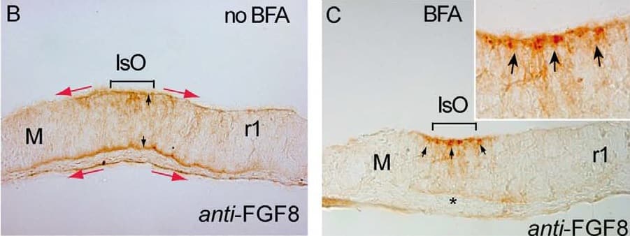
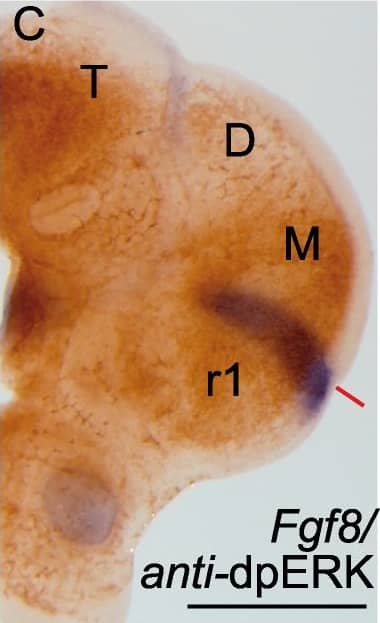
Bafilomycin A1 (BAF) treatment demonstrates the polarization of ERK activity by FGF8 signal activity along the neuraltube. A-C) BAF amplifies ectopic ERK1/2 activity-related FGF8 induction revealing a clear polarized distribution of ERK1/2 activity around FGF8soaked beads depending of the implanted bead; rostral to IsO (C,D,G), or caudal to IsO (C,E). Nonetheles, isthmic organizer morphogenetic activityseems unaffected for Fgf8 (A) and negative regulator Sprouty 2 (B) expressions. Note that PBS bead implantation in control side (blue asterisk in C andF) did not show any ectopic induction. Two hours after bead implantation a clear amplified and almost non-homogeneous ERK1/2 activity wasdetected rostrally in the mesencephalon (rostral to the IsO), which was detected caudally when bead was placed in hindbrain (caudal to the IsO)territories (E). In telencephalic vesicles, caudal to the anr (H) the polarity of ERK activation was reversed. This polarized dpERK detection around thebead is lost at the zli (zona limitans intrathalamica) region (I). Similar symmetric ERK-related FGF8 signal found in zli was seen when placing a FGF8bead in the midbrain of Fgf8 hypomorphic mice (J,K). Importantly FGF8b protein distribution (M) was observed apparently in equal intensity andrange at rostral (N) and caudal (O) sides of the bead (for comparison with PBS bead in panel L). Scale bars are 0,5 mm in A, B, C, H, I, 200 mm in D, E, J,100 mm in F, G, K, L, M, and 50 mm in N, O. Image collected and cropped by CiteAb from the following publication (https://www.ncbi.nlm.nih.gov/pmc/articles/PMC3391221/), licensed under a CC-BY license. Not internally tested by R&D Systems.Applications for Human/Mouse FGF-8 Antibody
Application
Recommended Usage
Immunohistochemistry
8-25 µg/mL
Sample: Immersion fixed paraffin-embedded sections of human prostate cancer tissue and normal human prostate
Sample: Immersion fixed paraffin-embedded sections of human prostate cancer tissue and normal human prostate
Western Blot
1 µg/mL
Sample: Recombinant Human FGF-8a (Catalog # 4745-F8)
Recombinant Mouse FG-8b (Catalog # 423-F8)
Recombinant Mouse FGF-8c (Catalog # 424-FC)
Recombinant Human FGF-8e (Catalog # 4746-F8)
Recombinant Human FGF-8f (Catalog # 5027-FF)
Sample: Recombinant Human FGF-8a (Catalog # 4745-F8)
Recombinant Mouse FG-8b (Catalog # 423-F8)
Recombinant Mouse FGF-8c (Catalog # 424-FC)
Recombinant Human FGF-8e (Catalog # 4746-F8)
Recombinant Human FGF-8f (Catalog # 5027-FF)
Neutralization
Measured by its ability to neutralize FGF-8-induced proliferation in the NR6R-3T3 mouse fibroblast cell line. Rizzino, A. et al. (1988) Cancer Res. 48:4266. The Neutralization Dose (ND50) is typically 0.25-0.75 µg/mL in the presence of 125 ng/mL Recombinant Mouse FGF-8 c Isoform and 0.1 µg/mL heparin.
Formulation, Preparation, and Storage
Purification
Protein A or G purified from ascites
Reconstitution
Reconstitute at 0.5 mg/mL in sterile PBS. For liquid material, refer to CoA for concentration.
Formulation
Lyophilized from a 0.2 μm filtered solution in PBS with Trehalose. *Small pack size (SP) is supplied either lyophilized or as a 0.2 µm filtered solution in PBS.
Shipping
Lyophilized product is shipped at ambient temperature. Liquid small pack size (-SP) is shipped with polar packs. Upon receipt, store immediately at the temperature recommended below.
Stability & Storage
Use a manual defrost freezer and avoid repeated freeze-thaw cycles.
- 12 months from date of receipt, -20 to -70 °C as supplied.
- 1 month, 2 to 8 °C under sterile conditions after reconstitution.
- 6 months, -20 to -70 °C under sterile conditions after reconstitution.
Background: FGF-8
References
- Mattila, M.M. and P.L. Harkonen (2007) Cytokine Growth Factor Rev. 18:257.
- Reuss, B. and O. von Bohlen und Halbach (2003) Cell Tiss. Res. 313:139.
- Tanaka, A. et al. (1992) Proc. Natl. Acad. Sci. USA 89:8928.
- Gemel, J. et al. (1996) Genomics 35:253.
- Olsen, S.K. et al. (2006) Genes Dev. 20:185.
- Crossley, P.H. et al. (1996) Cell, 84:127.
- Heikinheimo, M. et al. (1994) Mech. Dev. 48:129.
- Sun, X. et al. (1999) Genes Dev. 13:1834.
- Ghosh, A.K. et al. (1996) Cell Growth Differ. 7:1425.
- Mattila, M.M. et al. (2001) Oncogene 20:2791.
- Valve, E. et al. (2000) Int. J. Cancer 88:718.
- Valve, E.M. et al. (2001) Lab. Invest. 81:815.
- Nezu, M. et al. (2005) Biochem. Biophys. Res. Commun. 335:843.
Long Name
Fibroblast Growth Factor 8
Alternate Names
AIGF, FGF8, HBGF-8
Gene Symbol
FGF8
UniProt
Additional FGF-8 Products
Product Documents for Human/Mouse FGF-8 Antibody
Product Specific Notices for Human/Mouse FGF-8 Antibody
For research use only
Loading...
Loading...
Loading...
Loading...


