Human/Mouse Peroxiredoxin 3 Antibody
R&D Systems, part of Bio-Techne | Catalog # AF6610

Key Product Details
Species Reactivity
Validated:
Cited:
Applications
Validated:
Cited:
Label
Antibody Source
Product Specifications
Immunogen
Met1-Gln257
Accession # P20108
Specificity
Clonality
Host
Isotype
Scientific Data Images for Human/Mouse Peroxiredoxin 3 Antibody
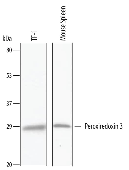
Detection of Human and Mouse Peroxiredoxin 3 by Western Blot.
Western blot shows lysates of TF-1 human erythroleukemic cell line and mouse spleen tissue. PVDF Membrane was probed with 1 µg/mL of Human/Mouse Peroxiredoxin 3 Antigen Affinity-purified Polyclonal Antibody (Catalog # AF6610) followed by HRP-conjugated Anti-Goat IgG Secondary Antibody (Catalog # HAF019). A specific band was detected for Peroxiredoxin 3 at approximately 28 kDa (as indicated). This experiment was conducted under reducing conditions and using Immunoblot Buffer Group 8.Detection of Mouse Peroxiredoxin 3 by Western Blot
Pharmacologic inhibition or genetic ablation of NOX reduces mitochondrial superoxide production in isolated CMs cultured in the presence of EtOH and ACA.(A) Scheme depicting incubation and imaging of isolated adult CMs from B6, ALDH2−/− or ALDH2−/−/gp91phox−/− mice with apocynin (300 μM), EtOH (100 μM) or ACA (100 μM). (B) MitoSOX-enhanced fluorescence per CMs. Quantification of 205–931 CMs on 6–12 independent low power fields per group. (C) Representative fluorescence images of CMs loaded with MitoSOX after culture for 18h in control media (leftmost column) in the presence of ACA 100 μM (middle column) and in the presence of ACA (100 μM) together with the NOX inhibitor apocynin (300 μM) rightmost column. (D) Peroxiredoxin 3 ratio monomer/dimer determined by Western blot under non-reducing, denaturing conditions in n = 4–7 independent samples per group. (E) Representative original Western blots. Image collected and cropped by CiteAb from the following publication (https://www.nature.com/articles/srep32554), licensed under a CC-BY license. Not internally tested by R&D Systems.Applications for Human/Mouse Peroxiredoxin 3 Antibody
Western Blot
Sample: TF‑1 human erythroleukemic cell line and mouse spleen tissue
Formulation, Preparation, and Storage
Purification
Reconstitution
Formulation
Shipping
Stability & Storage
- 12 months from date of receipt, -20 to -70 °C as supplied.
- 1 month, 2 to 8 °C under sterile conditions after reconstitution.
- 6 months, -20 to -70 °C under sterile conditions after reconstitution.
Background: Peroxiredoxin 3
Peroxiredoxin-3 (Prx-3/III; also AOP-1, MER5 and thioredoxin-dependent peroxidase reductase) is a ubiquitous, 22-28 kDa mitochondrial antioxidant enzyme that belongs to the 2-Cys class of the TSA/ahpC family of peroxiredoxins. Prx-3 is known to act as either a homodimer, or a decamer, and scavenge reactive oxygen species generated by oxidative stress. The mouse Prx-3 precursor molecule is 257 amino acids (aa) in length. It contains a cleavable N-terminal 63 aa mitochondrial targeting sequence, plus a 194 aa mature enzyme that shows a thioredoxin domain between aa 64-222. There are two catalytic cysteines, one at Cys109 and another at Cys230 of the precursor. Prx-3 undergoes a phosphorylation at Thr147 that reduces its activity. One potential splice form shows a deletion of Arg149, Lys150 and Arg185. Full-length mouse Prx-3 is 86% and 95% aa identical to human and rat Prx-3, respectively.
Alternate Names
Gene Symbol
UniProt
Additional Peroxiredoxin 3 Products
Product Documents for Human/Mouse Peroxiredoxin 3 Antibody
Product Specific Notices for Human/Mouse Peroxiredoxin 3 Antibody
For research use only

