Human/Mouse Phospho- Histone H2AX (S139) Antibody
R&D Systems, part of Bio-Techne | Catalog # 4418-APC-100


Discontinued Product
4418-APC-100 has been discontinued.
An alternative/replacement product is available:
AF2288. View all Histone H2AX products.
Key Product Details
Species Reactivity
Validated:
Human, Mouse
Cited:
Mouse, Rat, Plant - Brassica Rapa
Applications
Validated:
Immunocytochemistry, Western Blot
Cited:
Immunocytochemistry, Immunohistochemistry
Label
Unconjugated
Antibody Source
Polyclonal Rabbit IgG
Product Specifications
Immunogen
Phosphopeptide containing human Histone H2AX S139 site
Specificity
Detects human and mouse Histone H2AX when phosphorylated at S139 in Western blots. This antibody detects only double-strand DNA breaks.
Clonality
Polyclonal
Host
Rabbit
Isotype
IgG
Scientific Data Images for Human/Mouse Phospho- Histone H2AX (S139) Antibody
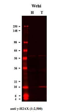
Detection of Phospho-Histone H2AX (S139) in Western blot.
Wehi cells were treated with (T)and without (H) 25 μM etoposide for 4 hours.Cells were lysed in Tris-Glycine SDS samplebuffer at the concentration 1 x 10⁷cells/ml and10 μl of each lysate were loaded per well of 4-20% Tris-Glycine gel. Proteins were transferredonto an Immobilon FL membrane and Phospho-H2AX (S139)was detected with anti-phosphorylated HistoneH2AX antibody (Cat# 4418-APC-020, 1:2500) followed by IR800-conjugated anti-rabbit secondary antibody.Phospho-Histone H2AX (S139) in C2C12 Mouse Cell Line.
Phospho-Histone H2AX (S139) was detected in immersion fixed C2C12 mouse myoblast cell line stimulated with etoposide (positive, left panel) and C2C12 mouse myoblast cell line (negative, right panel) using Rabbit Anti-Human/Mouse Phospho- Histone H2AX (S139) Antigen Affinity-purified Polyclonal Antibody (Catalog # 4418-APC-020) at 1.7 µg/mL for 3 hours at room temperature. Cells were stained using the NorthernLights™ 557-conjugated Anti-Rabbit IgG Secondary Antibody (red; NL004) and counterstained with DAPI (blue). Specific staining was localized to nuclei. Staining was performed using our protocol for Fluorescent ICC Staining of Non-adherent Cells.Applications for Human/Mouse Phospho- Histone H2AX (S139) Antibody
Application
Recommended Usage
Immunocytochemistry
1.7-15 µg/mL
Sample: mmersion fixed C2C12 mouse myoblast cell line stimulated with etoposide
Sample: mmersion fixed C2C12 mouse myoblast cell line stimulated with etoposide
Western Blot
1:2500 dilution
Sample: Wehi cells were treated with etoposide
Sample: Wehi cells were treated with etoposide
Formulation, Preparation, and Storage
Purification
Antigen Affinity-purified
Formulation
Supplied as a solution containing PBS.
Shipping
The product is shipped with polar packs. Upon receipt, store it immediately at the temperature recommended below.
Stability & Storage
Use a manual defrost freezer and avoid repeated freeze-thaw cycles.
- 12 months from date of receipt, -20 to -70 °C, as supplied.
- 1 month, 2 to 8 °C under sterile conditions after opening.
- 6 months, -20 to -70 °C under sterile conditions after opening.
Background: Histone H2AX
Additional Histone H2AX Products
Product Documents for Human/Mouse Phospho- Histone H2AX (S139) Antibody
Product Specific Notices for Human/Mouse Phospho- Histone H2AX (S139) Antibody
For research use only
Loading...
Loading...
Loading...
