Human/Mouse PTEN Antibody
R&D Systems, part of Bio-Techne | Catalog # MAB847R


Key Product Details
Species Reactivity
Applications
Label
Antibody Source
Product Specifications
Immunogen
Thr2-Val403
Accession # P60484
Specificity
Clonality
Host
Isotype
Scientific Data Images for Human/Mouse PTEN Antibody
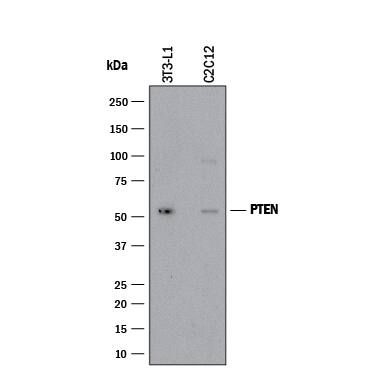
Detection of Mouse PTEN by Western Blot.
Western blot shows lysates of 3T3-L1 mouse embryonic fibroblast adipose-like cell line and C2C12 mouse myoblast cell line. PVDF membrane was probed with 2 µg/mL of Mouse Anti-Human/Mouse PTEN Monoclonal Antibody (Catalog # MAB847R) followed by HRP-conjugated Anti-Mouse IgG Secondary Antibody (HAF018). A specific band was detected for PTEN at approximately 54 kDa (as indicated). This experiment was conducted under reducing conditions and using Western Blot Buffer Group 1.Detection of Mouse PTEN by Western Blot.
Western blot shows lysates of mouse brain. PVDF membrane was probed with 2 µg/mL of Mouse Anti-Human/Mouse PTEN Monoclonal Antibody (Catalog # MAB847R) followed by HRP-conjugated Anti-Mouse IgG Secondary Antibody (HAF018). A specific band was detected for PTEN at approximately 54 kDa (as indicated). This experiment was conducted under reducing conditions and using Western Blot Buffer Group 1.Detection of PTEN in Human U937 cell line by Flow Cytometry.
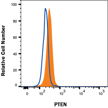
Human U937 histiocytic lymphoma cell line was stained with Mouse Anti-Human/Mouse PTEN Monoclonal Antibody (Catalog # MAB847R, filled histogram) or isotype control antibody (MAB002, open histogram), followed by Phycoerythrin-conjugated Anti-Mouse IgG F(ab')2 Secondary Antibody (F0102B). To facilitate intracellular staining, cells were fixed with Flow Cytometry Fixation Buffer (FC004) and permeabilized with Flow Cytometry Permeabilization/Wash Buffer I (FC005). Staining was performed using our protocol for Staining Intracellular Molecules.Applications for Human/Mouse PTEN Antibody
Intracellular Staining by Flow Cytometry
Sample: Human U937 histiocytic lymphoma cell line
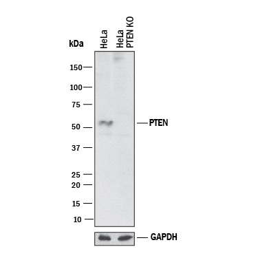
Knockout Validated
Western Blot
Sample: 3T3‑L1 mouse embryonic fibroblast adipose-like cell line, C2C12 mouse myoblast cell line and mouse brain
Formulation, Preparation, and Storage
Purification
Reconstitution
Formulation
Shipping
Stability & Storage
- 12 months from date of receipt, -20 to -70 °C as supplied.
- 1 month, 2 to 8 °C under sterile conditions after reconstitution.
- 6 months, -20 to -70 °C under sterile conditions after reconstitution.
Background: PTEN
The tumor suppressor gene PTEN (phosphatase and tensin homolog deleted on chromosome 10), also known as MMAC1 (mutated in multiple advanced cancers 1), encodes a phosphatase that contains the catalytic signature motif (HCxxGxxRS/T) found in all members of the protein tyrosine phosphatase family. In vitro, the recombinant PTEN has both lipid phosphatase and protein phosphatase activities (1, 2). Interestingly, accumulating evidence has shown that the tumor suppressor activity of PTEN relies on its ability to dephosphorylate phosphatidylinositol (3,4,5)-triphosphate specifically at position 3 of the inositol ring (3). This activity reduces the levels of phosphatidylinositol (3,4,5)-triphosphate which is specifically produced from phosphatidylinositol (4,5)-diphosphate by PI 3-kinase upon activation by a variety of stimuli. Therefore, PTEN antagonizes PI 3-kinase-induced downstream signaling events and cellular processes including cell growth, apoptosis and cell motility. In vivo, the importance of PTEN catalytic activity in its tumor suppressor functions is underscored by the fact that the majority of PTEN missense mutations detected in tumor specimens target the phosphatase domain and cause a loss in PTEN phosphatase activity (4).
References
- Maehama, T. and J. Dixon (1998) J. Biol. Chem. 273:13375.
- Das, S. et al. (2003) Proc. Natl. Acad. Sci. USA 100:7491.
- Myers, M. et al. (1998) Proc. Natl. Acad. Sci. USA 95:13513.
- Waite, K. and C. Eng (2002) Am. J. Hum. Genet. 70:829.
Long Name
Alternate Names
Gene Symbol
UniProt
Additional PTEN Products
Product Documents for Human/Mouse PTEN Antibody
Product Specific Notices for Human/Mouse PTEN Antibody
For research use only


