Human/Mouse/Rat alpha-Actinin 1 Antibody
R&D Systems, part of Bio-Techne | Catalog # MAB8279

Key Product Details
Validated by
Species Reactivity
Validated:
Cited:
Applications
Validated:
Cited:
Label
Antibody Source
Product Specifications
Immunogen
Asn272-Thr500
Accession # P12814
Specificity
Clonality
Host
Isotype
Scientific Data Images for Human/Mouse/Rat alpha-Actinin 1 Antibody
Detection of Human, Mouse, and Rat alpha-Actinin 1 by Western Blot.
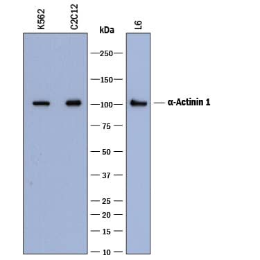
Western blot shows lysates of K562 human chronic myelogenous leukemia cell line, C2C12 mouse myoblast cell line, and L6 rat myoblast cell line. PVDF membrane was probed with 0.2 µg/mL of Mouse Anti-Human/Mouse/Rat a-Actinin 1 Monoclonal Antibody (Catalog # MAB8279) followed by HRP-conjugated Anti-Mouse IgG Secondary Antibody (Catalog # HAF018). A specific band was detected for a-Actinin 1 at approximately 100-105 kDa (as indicated). This experiment was conducted under reducing conditions and using Immunoblot Buffer Group 1.Detection of Mouse alpha-Actinin 1 by Simple WesternTM.
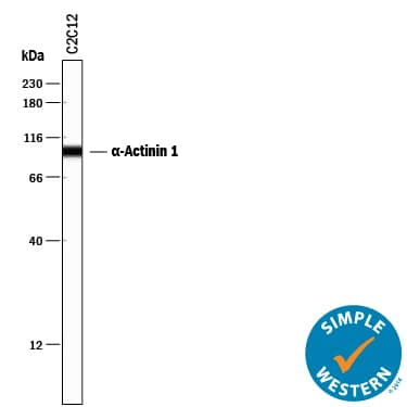
Simple Western lane view shows lysates of C2C12 mouse myoblast cell line, loaded at 0.5 mg/mL. A specific band was detected for alpha-Actinin 1 at approximately 98 kDa (as indicated) using 2 µg/mL of Mouse Anti-Human/Mouse/Rat alpha-Actinin 1 Monoclonal Antibody (Catalog # MAB8279). This experiment was conducted under reducing conditions and using the 12-230 kDa separation system.Detection of Human alpha-Actinin 1 by Simple Western
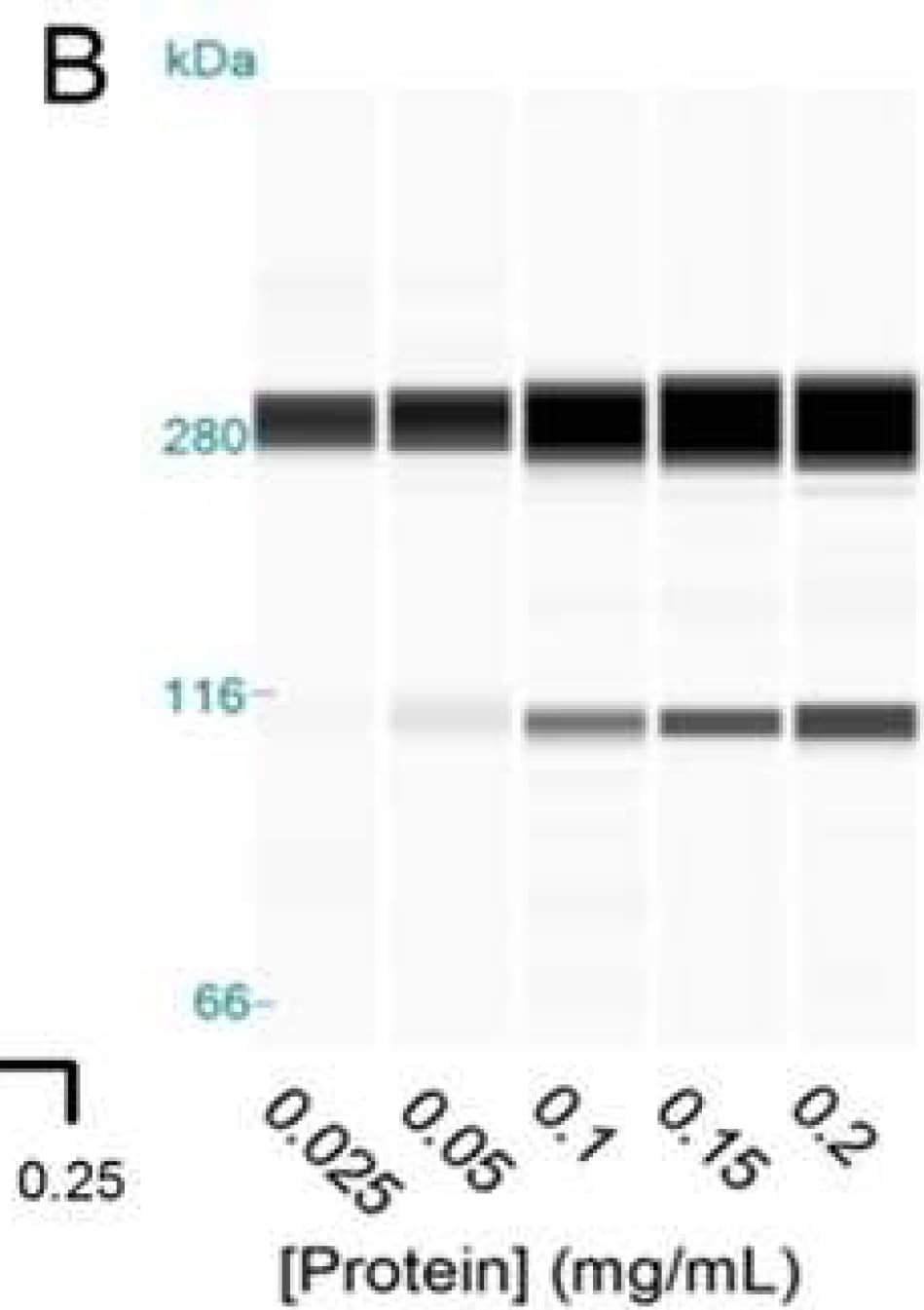
Detection and quantification of SI protein. (Panels A) and (Panels B) show the concentration-dependence of SI and alpha-actinin signals (Panel C) using automated capillary Western blotting of Caco-2/TC7 lysates. (Panel D) shows Caco-2/TC7 cell surface protein fraction purified after biotinylation from cells cultured on 6-well Transwell filters in glucose or sucrose for 21 days and treated on the apical side for the final 3 days with 1.5 mg/mL OLE. (Panel E) shows the total cellular SI, relative to alpha-actinin with data normalised to the glucose control and presented as mean ± SEM with n = 12, analysed in duplicate runs. The size was determined relative to a protein standard ladder for SI in the total lysate relative to alpha-actinin (F), normalised and presented as mean ± SEM with n = 24. * p < 0.05, ** p < 0.01, *** p < 0.001 from ANOVA followed by Tukey’s post-hoc test. There was no change in the alpha-actinin loading control between the groups. Image collected and cropped by CiteAb from the following publication (https://pubmed.ncbi.nlm.nih.gov/31266155), licensed under a CC-BY license. Not internally tested by R&D Systems.Applications for Human/Mouse/Rat alpha-Actinin 1 Antibody
Simple Western
Sample: C2C12 mouse myoblast cell line
Western Blot
Sample: K562 human chronic myelogenous leukemia cell line, C2C12 mouse myoblast cell line, and L6 rat myoblast cell line
Formulation, Preparation, and Storage
Purification
Reconstitution
Formulation
Shipping
Stability & Storage
- 12 months from date of receipt, -20 to -70 °C as supplied.
- 1 month, 2 to 8 °C under sterile conditions after reconstitution.
- 6 months, -20 to -70 °C under sterile conditions after reconstitution.
Background: alpha Actinin 1
alpha-Actinin 1 (ACTN1) is a member of the spectrin superfamily with a predicted molecular weight of approximately 100 kDa. It is 892 amino acids (aa) in length and shares 99% aa identity with mouse and rat ACTN1. Multiple splice forms exist, resulting in 4 distinct isoforms with different tissue expression patterns. ACTN1 binds to F-actin as well as other cytoskeletal and signaling proteins and plays an important role in both cell motility and muscle contraction.
Long Name
Alternate Names
Gene Symbol
UniProt
Additional alpha Actinin 1 Products
Product Documents for Human/Mouse/Rat alpha-Actinin 1 Antibody
Product Specific Notices for Human/Mouse/Rat alpha-Actinin 1 Antibody
For research use only


