Human/Mouse/Rat Annexin A1 Antibody
R&D Systems, part of Bio-Techne | Catalog # MAB37701

Key Product Details
Validated by
Species Reactivity
Validated:
Cited:
Applications
Validated:
Cited:
Label
Antibody Source
Product Specifications
Immunogen
Met1-Asn346
Accession # P04083
Specificity
Clonality
Host
Isotype
Scientific Data Images for Human/Mouse/Rat Annexin A1 Antibody
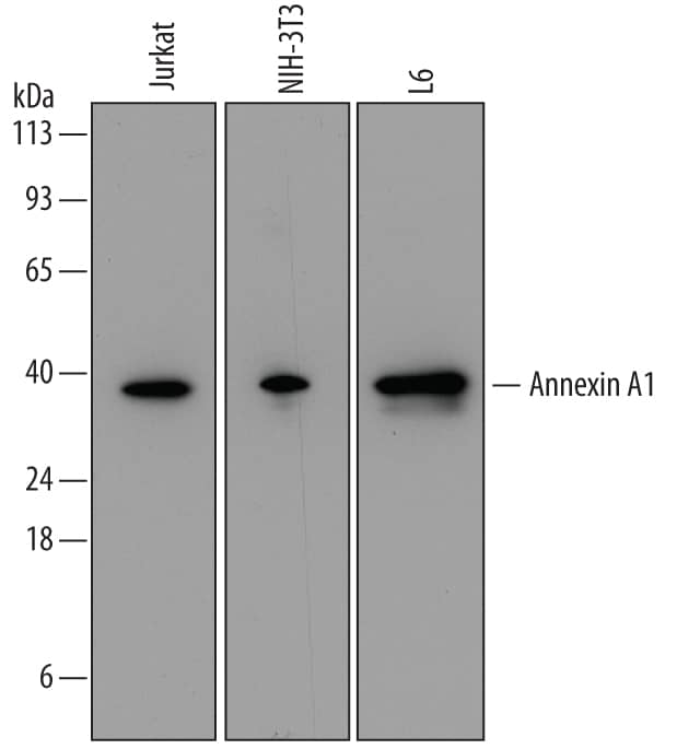
Detection of Human, Mouse, and Rat Annexin A1 by Western Blot.
Western blot shows lysates of Jurkat human acute T cell leukemia cell line, NIH-3T3 mouse embryonic fibroblast cell line, and L6 rat myoblast cell line. PVDF membrane was probed with 0.1 µg/mL of Mouse Anti-Human/Mouse Annexin A1 Monoclonal Antibody (Catalog # MAB37701) followed by HRP-conjugated Anti-Mouse IgG Secondary Antibody (Catalog # HAF007). A specific band was detected for Annexin A1 at approximately 39 kDa (as indicated). This experiment was conducted under reducing conditions and using Immunoblot Buffer Group 2.Detection of Human Annexin A1 by Simple WesternTM.
Simple Western lane view shows lysates of Jurkat human acute T cell leukemia cell line, loaded at 0.2 mg/mL. A specific band was detected for Annexin A1 at approximately 45 kDa (as indicated) using 1 µg/mL of Mouse Anti-Human/Mouse/Rat Annexin A1 Monoclonal Antibody (Catalog # MAB37701). This experiment was conducted under reducing conditions and using the 12-230 kDa separation system.Detection of Mouse Annexin A1 by Simple WesternTM.
Simple Western lane view shows lysates of NIH‑3T3 mouse embryonic fibroblast cell line, loaded at 0.2 mg/mL. A specific band was detected for Annexin A1 at approximately 45 kDa (as indicated) using 1 µg/mL of Mouse Anti-Human/Mouse/Rat Annexin A1 Monoclonal Antibody (Catalog # MAB37701). This experiment was conducted under reducing conditions and using the 12-230 kDa separation system.Applications for Human/Mouse/Rat Annexin A1 Antibody
Knockout Validated
Simple Western
Sample: Jurkat human acute T cell leukemia cell line and NIH‑3T3 mouse embryonic fibroblast cell line
Western Blot
Sample: Jurkat human acute T cell leukemia cell line, NIH‑3T3 mouse embryonic fibroblast cell line, and L6 rat myoblast cell line.
Formulation, Preparation, and Storage
Purification
Reconstitution
Formulation
Shipping
Stability & Storage
- 12 months from date of receipt, -20 to -70 °C as supplied.
- 1 month, 2 to 8 °C under sterile conditions after reconstitution.
- 6 months, -20 to -70 °C under sterile conditions after reconstitution.
Background: Annexin A1
The Annexins are a family of Calcium-dependent phospholipid-binding proteins that are preferentially located on the cytosolic face of the plasma membrane. The Annexins have a molecular weight of approximately 35 to 40 kDa and consist of a unique amino terminal domain followed by a homologous C-terminal core domain containing the calcium-dependent phospholipid-binding sites. The C‑terminal domain is comprised of four 60‑70 amino acid repeats, known as annexin repeats or an endonexin fold (Annexin A6 contains 8 annexin repeats). The four annexin repeats form a highly alpha-helical, tightly packed disc known as the annexin domain, which binds to phospholipids in the membrane in a calcium-dependent manner. Members of the annexin family play a role in cytoskeletal interactions, phospholipase inhibition, regulation of cellular growth, and intracellular signal transduction pathways. Annexin A1 (ANXA1), also known as annexin I, lipocortin I, and calpactin II, is an ~ 40 kDa protein with phospholipase A2 inhibitory activity. Since phospholipase A2 is required for the biosynthesis of the potent mediators of inflammation, prostaglandins and leukotrienes, Annexin A1 may have anti-inflammatory activity. Human Annexin A1 shares 88 and 89% amino acid sequence identity with mouse and rat Annexin A1, respectively.
Alternate Names
Gene Symbol
UniProt
Additional Annexin A1 Products
Product Documents for Human/Mouse/Rat Annexin A1 Antibody
Product Specific Notices for Human/Mouse/Rat Annexin A1 Antibody
For research use only


