Human/Mouse/Rat CDC25B Antibody
R&D Systems, part of Bio-Techne | Catalog # AF1649

Key Product Details
Species Reactivity
Applications
Label
Antibody Source
Product Specifications
Immunogen
Glu391-Gln580
Accession # P30305
Specificity
Clonality
Host
Isotype
Scientific Data Images for Human/Mouse/Rat CDC25B Antibody
Detection of Human/Mouse/Rat CDC25B by Western Blot.
Western blot shows lysates of HeLa human cervical epithelial carcinoma cell line, DA3 mouse myeloma cell line, and Nb2-11 rat lymphoma cell line. PVDF membrane was probed with 0.3 µg/mL of Goat Anti-Human/Mouse/Rat CDC25B Antigen Affinity-purified Polyclonal Antibody (Catalog # AF1649) followed by HRP-conjugated Anti-Goat IgG Secondary Antibody (Catalog # HAF017). A specific band was detected for CDC25B at approximately 61 - 67 kDa (as indicated). This experiment was conducted under reducing conditions and using Immunoblot Buffer Group 1.CDC25B in HL‑60 Human Cell Line.
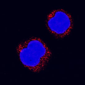
CDC25B was detected in immersion fixed HL-60 human acute promyelocytic leukemia cell line using Goat Anti-Human/Mouse/Rat CDC25B Antigen Affinity-purified Polyclonal Antibody (Catalog # AF1649) at 15 µg/mL for 3 hours at room temperature. Cells were stained using the NorthernLights™ 557-conjugated Anti-Goat IgG Secondary Antibody (red; Catalog # NL001) and counterstained with DAPI (blue). Specific staining was localized to cytoplasm. View our protocol for Fluorescent ICC Staining of Non-adherent Cells.Applications for Human/Mouse/Rat CDC25B Antibody
Immunocytochemistry
Sample: Immersion fixed human peripheral blood mononuclear cells and HL‑60 human acute promyelocytic leukemia cell line
Western Blot
Sample: HeLa human cervical epithelial carcinoma cell line, DA3 mouse myeloma cell line, and Nb2-11 rat lymphoma cell line
Reviewed Applications
Read 1 review rated 5 using AF1649 in the following applications:
Formulation, Preparation, and Storage
Purification
Reconstitution
Formulation
Shipping
Stability & Storage
- 12 months from date of receipt, -20 to -70 °C as supplied.
- 1 month, 2 to 8 °C under sterile conditions after reconstitution.
- 6 months, -20 to -70 °C under sterile conditions after reconstitution.
Background: CDC25B
Cell Division Cycle 25B (Cdc25B) phosphatase removes inorganic phosphate groups covalently attached to tyrosine, serine and threonine residues in proteins (1). Breast cancer patients bearing tumors containing high levels of Cdc25B have been found to have a greater incidence of aggressive, high-grade tumors than those with low Cdc25B levels (2). In cells, the levels of Cdc25B activity are highest during the G2/M transition of the cell cycle, where it is suspected to be involved in “checkpoint” control of cell cycle progression (3). Overexpression of Cdc25B reduces the G2/M cell cycle block caused by ionizing radiation (4). Although activated by phosphorylation, Ser323 phosphorylation causes the enzyme to bind the protein 14-3-3, preventing substrate access to the catalytic site (5). One of the major substrates of Cdc25B is Cdc2, a kinase that is activated by dephosphorylation (6). The recombinant protein is truncated to remove the N‑terminal regulatory domains and is fully active.
References
- Draetta, G. and J. Eckstein (1997) Biochim. Biophys. Acta 1332:M53.
- Galaktionov, K. et al. (1995) Science 269:1575.
- Lammer, C. et al. (1998) J. Cell Sci. 111:2445.
- Miyata, H. et al. (2001) Cancer Res. 61:3188.
- Forrest, A. and B. Gabrielli (2001) Oncogene 20:4393.
- Gautier, J. et al. (1991) Cell 67:197.
Long Name
Alternate Names
Entrez Gene IDs
Gene Symbol
UniProt
Additional CDC25B Products
Product Documents for Human/Mouse/Rat CDC25B Antibody
Product Specific Notices for Human/Mouse/Rat CDC25B Antibody
For research use only

