Human/Mouse/Rat Complexin-1/2 Antibody
R&D Systems, part of Bio-Techne | Catalog # MAB8069

Key Product Details
Species Reactivity
Human, Mouse, Rat
Applications
Immunohistochemistry, Simple Western, Western Blot
Label
Unconjugated
Antibody Source
Monoclonal Mouse IgG1 Clone # 837802
Product Specifications
Immunogen
E. coli-derived recombinant human Complexin-1
Met1-Lys134
Accession # O14810
Met1-Lys134
Accession # O14810
Specificity
Detects human Complexin-1 and Complexin-2 in ELISAs. Detects human, mouse, and rat Complexin-1 and -2 in Western blots.
Clonality
Monoclonal
Host
Mouse
Isotype
IgG1
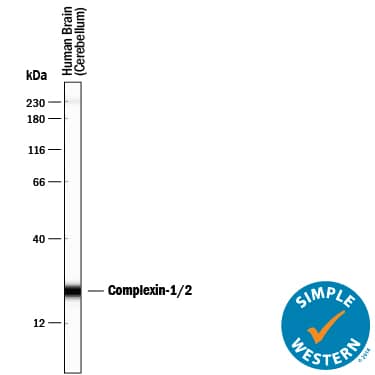
Scientific Data Images for Human/Mouse/Rat Complexin-1/2 Antibody
Detection of Human, Mouse, and Rat Complexin-1/2 by Western Blot.
Western blot shows lysates of human brain (cerebellum) tissue, mouse brain (cortex) tissue, and rat brain (cerebellum) tissue. PVDF membrane was probed with 0.5 µg/mL of Mouse Anti-Human/Mouse/Rat Complexin-1/2 Monoclonal Antibody (Catalog # MAB8069) followed by HRP-conjugated Anti-Mouse IgG Secondary Antibody (Catalog # HAF018). Specific bands were detected for Complexin-1/2 at approximately 17-19 kDa (as indicated). This experiment was conducted under reducing conditions and using Immunoblot Buffer Group 1.Complexin-1/2 in Human Pancreas.
Complexin-1/2 was detected in immersion fixed paraffin-embedded sections of human pancreas using Mouse Anti-Human/Mouse/Rat Complexin-1/2 Monoclonal Antibody (Catalog # MAB8069) at 15 µg/mL overnight at 4 °C. Before incubation with the primary antibody, tissue was subjected to heat-induced epitope retrieval using Antigen Retrieval Reagent-Basic (Catalog # CTS013). Tissue was stained using the Anti-Mouse HRP-DAB Cell & Tissue Staining Kit (brown; Catalog # CTS002) and counterstained with hematoxylin (blue). Specific staining was localized to islets. View our protocol for Chromogenic IHC Staining of Paraffin-embedded Tissue Sections.Detection of Human Complexin-1/2 by Simple WesternTM.
Simple Western lane view shows lysates of human brain (cerebellum) tissue, loaded at 0.5 mg/mL. A specific band was detected for Complexin-1/2 at approximately 23 kDa (as indicated) using 5 µg/mL of Mouse Anti-Human/Mouse/Rat Complexin-1/2 Monoclonal Antibody (Catalog # MAB8069). This experiment was conducted under reducing conditions and using the 12-230 kDa separation system. Non-specific interaction with the 230 kDa Simple Western standard may be seen with this antibody.Applications for Human/Mouse/Rat Complexin-1/2 Antibody
Application
Recommended Usage
Immunohistochemistry
8-25 µg/mL
Sample: Immersion fixed paraffin-embedded sections of human pancreas
Sample: Immersion fixed paraffin-embedded sections of human pancreas
Simple Western
5 µg/mL
Sample: Human brain (cerebellum) tissue
Sample: Human brain (cerebellum) tissue
Western Blot
0.5 µg/mL
Sample: Human brain (cerebellum) tissue, mouse brain (cortex) tissue, and rat brain (cerebellum) tissue
Sample: Human brain (cerebellum) tissue, mouse brain (cortex) tissue, and rat brain (cerebellum) tissue
Formulation, Preparation, and Storage
Purification
Protein A or G purified from hybridoma culture supernatant
Reconstitution
Reconstitute at 0.5 mg/mL in sterile PBS. For liquid material, refer to CoA for concentration.
Formulation
Lyophilized from a 0.2 μm filtered solution in PBS and NaCl with Trehalose. *Small pack size (SP) is supplied either lyophilized or as a 0.2 µm filtered solution in PBS.
Shipping
Lyophilized product is shipped at ambient temperature. Liquid small pack size (-SP) is shipped with polar packs. Upon receipt, store immediately at the temperature recommended below.
Stability & Storage
Use a manual defrost freezer and avoid repeated freeze-thaw cycles.
- 12 months from date of receipt, -20 to -70 °C as supplied.
- 1 month, 2 to 8 °C under sterile conditions after reconstitution.
- 6 months, -20 to -70 °C under sterile conditions after reconstitution.
Background: Complexin-1/2
Alternate Names
CPLX1;CPX1;CPX-I;DEE63;EIEE63
UniProt
Additional Complexin-1/2 Products
Product Documents for Human/Mouse/Rat Complexin-1/2 Antibody
Product Specific Notices for Human/Mouse/Rat Complexin-1/2 Antibody
For research use only
Loading...
Loading...
Loading...
Loading...


