Human/Mouse/Rat MuRF1/TRIM63 Antibody
R&D Systems, part of Bio-Techne | Catalog # AF5366

Key Product Details
Validated by
Species Reactivity
Validated:
Cited:
Applications
Validated:
Cited:
Label
Antibody Source
Product Specifications
Immunogen
Met1-Gly325
Accession # Q969Q1
Specificity
Clonality
Host
Isotype
Scientific Data Images for Human/Mouse/Rat MuRF1/TRIM63 Antibody
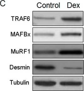
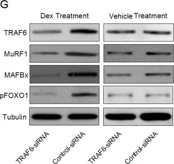
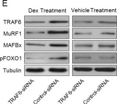
Detection of Human/Mouse/Rat MuRF1 by Western Blot.
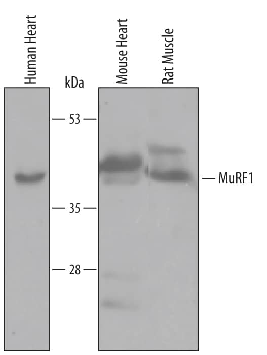
Western blot shows lysates of human heart, mouse heart, and rat muscle tissue. PVDF membrane was probed with 1 µg/mL of Goat Anti-Human/Mouse/Rat MuRF1 Antigen Affinity-purified Polyclonal Antibody (Catalog # AF5366) followed by HRP-conjugated Anti-Goat IgG Secondary Antibody (Catalog # HAF019). A specific band was detected for MuRF1 at approximately 41-44 kDa (as indicated). This experiment was conducted under reducing conditions and using Immunoblot Buffer Group 8.Detection of Human MuRF1/TRIM63 by Simple WesternTM.
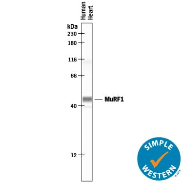
Simple Western lane view shows lysates of human heart tissue, loaded at 0.2 mg/mL. A specific band was detected for MuRF1/TRIM63 at approximately 46 kDa (as indicated) using 10 µg/mL of Goat Anti-Human/Mouse/Rat MuRF1/TRIM63 Antigen Affinity-purified Polyclonal Antibody (Catalog # AF5366) followed by 1:50 dilution of HRP-conjugated Anti-Goat IgG Secondary Antibody (Catalog # HAF109). This experiment was conducted under reducing conditions and using the 12-230 kDa separation system.Detection of Zebrafish MuRF1/TRIM63 by Western Blot
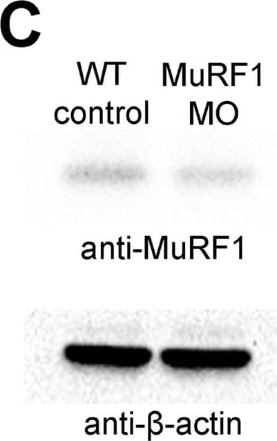
Blocking MuRF1-mediated proteasome degradation preserves myofibril integrity in tre/ncx1 deficient hearts.(A) Z-lines were visualized by alpha-actinin staining. By 72 hpf, sarcomeres are disassembled in hearts of uninjected (tre) and control morpholino-injected (tre +ctlMO) tremblor embryos. Murf1a/murf1 b knockdown does not affect sarcomere integrity in wild type embryos (WT +MO), but prevents sarcomere degradation in tre (tre +MO). Similarly, treatment with a proteasome inhibitor, MG132, preserves myofibril integrity in tre cardiomyocytes (tre +MG132). Scale bar, 10 μm. (B) Graph shows % of embryos with periodic alpha-actinin staining at 72 hpf. (C) Western blot detecting MuRF1 and beta-actin proteins in uninjected control (WT control) and murf1a/murf1 b knockdown (MuRF1 MO) embryos. Chi-squared test, *p<0.05; **p<0.01; ***p<0.001. Image collected and cropped by CiteAb from the following publication (https://pubmed.ncbi.nlm.nih.gov/28826496), licensed under a CC-BY license. Not internally tested by R&D Systems.Applications for Human/Mouse/Rat MuRF1/TRIM63 Antibody
Simple Western
Sample: Human heart tissue
Western Blot
Sample: Human heart, mouse heart, and rat muscle tissue
Formulation, Preparation, and Storage
Purification
Reconstitution
Formulation
Shipping
Stability & Storage
- 12 months from date of receipt, -20 to -70 °C as supplied.
- 1 month, 2 to 8 °C under sterile conditions after reconstitution.
- 6 months, -20 to -70 °C under sterile conditions after reconstitution.
Background: MuRF1/TRIM63
TRIM63 (Tripartite motif-containing protein 63; also MURF-1, SMRZ and RING finger protein 28) is a 41 kDa member of the RING finger-B-box-coiled-coil family of proteins. It is a striated muscle protein that is found in both cytoplasm and nucleus. TRIM63 has multiple finctions, among which are the inhibition of PKC epsilon-mediated cardiomyocyte hypertrophy and the maintenance of skeletal muscle M-line integrity. Human TRIM63 is 353 amino acids (aa) in length. It contains one RING finger domain (aa 23‑82), a B-Box type zinc-finger region (aa 117‑159), a coiled-coil region (aa 207‑269) and a C-terminal COS domain. Isoforms of TRIM63 show one potential alternate start site at Met14, a deletion of aa 105‑132 and a 21 aa substitution for aa 326‑353. Over aa 1‑325, human TRIM63 exhibits 93% aa identity with mouse TRIM63.
Long Name
Alternate Names
Gene Symbol
UniProt
Additional MuRF1/TRIM63 Products
Product Documents for Human/Mouse/Rat MuRF1/TRIM63 Antibody
Product Specific Notices for Human/Mouse/Rat MuRF1/TRIM63 Antibody
For research use only
