Human/Mouse/Rat Thioredoxin Reductase 1/TRXR1 Antibody
R&D Systems, part of Bio-Techne | Catalog # MAB7428


Conjugate
Catalog #
Key Product Details
Species Reactivity
Validated:
Human, Mouse, Rat
Cited:
Human
Applications
Validated:
CyTOF-ready, Intracellular Staining by Flow Cytometry, Western Blot
Cited:
Western Blot
Label
Unconjugated
Antibody Source
Monoclonal Mouse IgG1 Clone # 489804
Product Specifications
Immunogen
E. coli-derived recombinant mouse Thioredoxin Reductase 1/TRXR1
Met1-Ile497
Accession # Q16881
Met1-Ile497
Accession # Q16881
Specificity
Detects mouse Thioredoxin Reductase 1/TRXR1 in direct ELISAs and human, mouse, and rat Thioredoxin Reductase 1/TRXR1 in Western blots.
Clonality
Monoclonal
Host
Mouse
Isotype
IgG1
Scientific Data Images for Human/Mouse/Rat Thioredoxin Reductase 1/TRXR1 Antibody
Detection of Human, Mouse, and Rat Thioredoxin Reductase 1/TRXR1 by Western Blot.
Western blot shows lysates of HeLa human cervical epithelial carcinoma cell line, NIH-3T3 mouse embryonic fibroblast cell line, and C6 rat glioma cell line. PVDF membrane was probed with 0.1 µg/mL of Mouse Anti-Human Thioredoxin Reductase 1/TRXR1 Monoclonal Antibody (Catalog # MAB7428) followed by HRP-conjugated Anti-Mouse IgG Secondary Antibody (Catalog # HAF007). A specific band was detected for Thioredoxin Reductase 1/TRXR1 at approximately 65 kDa (as indicated). This experiment was conducted under reducing conditions and using Immunoblot Buffer Group 1.Detection of Thioredoxin Reductase 1/TRXR1 in HeLa Human Cell Line by Flow Cytometry.
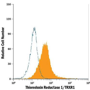
HeLa human cervical epithelial carcinoma cell line was stained with Mouse Anti-Human/Mouse/Rat Thioredoxin Reductase 1/TRXR1 Monoclonal Antibody (Catalog # MAB7428, filled histogram) or isotype control antibody (Catalog # MAB002, open histogram), followed by Allophycocyanin-conjugated Anti-Mouse IgG Secondary Antibody (Catalog # F0101B). To facilitate intracellular staining, cells were fixed with paraformaldehyde and permeabilized with saponin.Applications for Human/Mouse/Rat Thioredoxin Reductase 1/TRXR1 Antibody
Application
Recommended Usage
CyTOF-ready
Ready to be labeled using established conjugation methods. No BSA or other carrier proteins that could interfere with conjugation.
Intracellular Staining by Flow Cytometry
2.5 µg/106 cells
Sample: HeLa human cervical epithelial carcinoma cell line fixed with paraformaldehyde and permeabilized with saponin
Sample: HeLa human cervical epithelial carcinoma cell line fixed with paraformaldehyde and permeabilized with saponin
Western Blot
0.1 µg/mL
Sample: HeLa human cervical epithelial carcinoma cell line, NIH‑3T3 mouse embryonic fibroblast cell line, and C6 rat glioma cell line
Sample: HeLa human cervical epithelial carcinoma cell line, NIH‑3T3 mouse embryonic fibroblast cell line, and C6 rat glioma cell line
Reviewed Applications
Read 1 review rated 5 using MAB7428 in the following applications:
Formulation, Preparation, and Storage
Purification
Protein A or G purified from hybridoma culture supernatant
Reconstitution
Sterile PBS to a final concentration of 0.5 mg/mL. For liquid material, refer to CoA for concentration.
Formulation
Lyophilized from a 0.2 μm filtered solution in PBS with Trehalose. *Small pack size (SP) is supplied either lyophilized or as a 0.2 µm filtered solution in PBS.
Shipping
Lyophilized product is shipped at ambient temperature. Liquid small pack size (-SP) is shipped with polar packs. Upon receipt, store immediately at the temperature recommended below.
Stability & Storage
Use a manual defrost freezer and avoid repeated freeze-thaw cycles.
- 12 months from date of receipt, -20 to -70 °C as supplied.
- 1 month, 2 to 8 °C under sterile conditions after reconstitution.
- 6 months, -20 to -70 °C under sterile conditions after reconstitution.
Background: Thioredoxin Reductase 1/TRXR1
Alternate Names
GRIM-12, KDRF, TR1, TRXR1, TRXRD1, TXNR, TXNRD1
Gene Symbol
TXNRD1
UniProt
Additional Thioredoxin Reductase 1/TRXR1 Products
Product Documents for Human/Mouse/Rat Thioredoxin Reductase 1/TRXR1 Antibody
Product Specific Notices for Human/Mouse/Rat Thioredoxin Reductase 1/TRXR1 Antibody
For research use only
Loading...
Loading...
Loading...
Loading...
Loading...
