Human/Mouse RPTOR Antibody
R&D Systems, part of Bio-Techne | Catalog # MAB5957


Key Product Details
Species Reactivity
Applications
Label
Antibody Source
Product Specifications
Immunogen
Lys77-Gln230
Accession # Q8N122
Specificity
Clonality
Host
Isotype
Scientific Data Images for Human/Mouse RPTOR Antibody
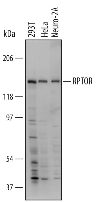
Detection of Human and Mouse RPTOR by Western Blot.
Western blot shows lysates of 293T human embryonic kidney cell line, HeLa human cervical epithelial carcinoma cell line, and Neuro-2A mouse neuroblastoma cell line. PVDF Membrane was probed with 1 µg/mL of Human/Mouse RPTOR Monoclonal Antibody (Catalog # MAB5957) followed by HRP-conjugated Anti-Mouse IgG Secondary Antibody (Catalog # HAF007). A specific band was detected for RPTOR at approximately 150 kDa (as indicated). This experiment was conducted under non-reducing conditions and using Immunoblot Buffer Group 1.Detection of RPTOR in HeLa Human Cell Line by Flow Cytometry.
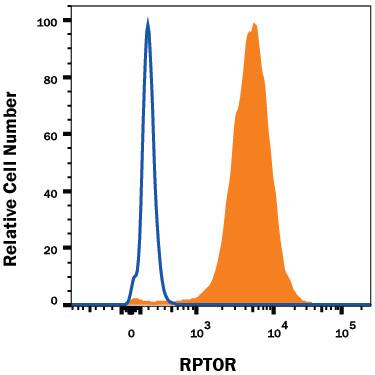
HeLa human cervical epithelial carcinoma cell line was stained with Mouse Anti-Human/Mouse RPTOR Monoclonal Antibody (Catalog # MAB5957, filled histogram) or isotype control antibody (Catalog # MAB0041, open histogram), followed by Phycoerythrin-conjugated Anti-Mouse IgG Secondary Antibody (Catalog # F0102B). To facilitate intracellular staining, cells were fixed with Flow Cytometry Fixation Buffer (Catalog # FC004) and permeabilized with Flow Cytometry Permeabilization/Wash Buffer I (Catalog # FC005). View our protocol for Staining Intracellular Molecules.Detection of RPTOR in C2C12 Mouse Cell Line by Flow Cytometry.
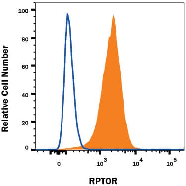
C2C12 mouse myoblast cell line was stained with Mouse Anti-Human/Mouse RPTOR Monoclonal Antibody (Catalog # MAB5957, filled histogram) or isotype control antibody (Catalog # MAB0041, open histogram), followed by Phycoerythrin-conjugated Anti-Mouse IgG Secondary Antibody (Catalog # F0102B). To facilitate intracellular staining, cells were fixed with Flow Cytometry Fixation Buffer (Catalog # FC004) and permeabilized with Flow Cytometry Permeabilization/Wash Buffer I (Catalog # FC005). View our protocol for Staining Intracellular Molecules.Applications for Human/Mouse RPTOR Antibody
CyTOF-ready
Intracellular Staining by Flow Cytometry
Sample: HeLa human cervical epithelial carcinoma cell line and C2C12 mouse myoblast cell line fixed with Flow Cytometry Fixation Buffer (Catalog # FC004) and permeabilized with Flow Cytometry Permeabilization/Wash Buffer I (Catalog # FC005)
Western Blot
Sample: 293T human embryonic kidney cell line, HeLa human cervical epithelial carcinoma cell line, and Neuro‑2A mouse neuroblastoma cell line
Reviewed Applications
Read 1 review rated 3 using MAB5957 in the following applications:
Formulation, Preparation, and Storage
Purification
Reconstitution
Formulation
Shipping
Stability & Storage
- 12 months from date of receipt, -20 to -70 °C as supplied.
- 1 month, 2 to 8 °C under sterile conditions after reconstitution.
- 6 months, -20 to -70 °C under sterile conditions after reconstitution.
Background: RPTOR
RPTOR (Raptor) is a 150 kDa component of the cytosolic mammalian target of Rapamycin complex 1 (mTORC1) which also contains mTOR and GBL proteins. mTORC1 plays a dominant role in cell cycle regulation in response to metabolic conditions. The interaction of RPTOR with the kinase mTOR is stabilized under conditions of nutrient deprivation and energy stress, leading to inhibition of mTOR and cell cycle arrest. RPTOR contains multiple Ser and Thr residues whose phosphorylation regulates the activation status of mTOR. RPTOR is critical for the response of skeletal muscle and adipose tissue to insulin. It contains three RNC blocks (aa 48-511), three HEAT repeats (aa 550-667), and seven C-terminal WD40 domains (aa 1020-1335). Within aa 77-230, human RPTOR shares 100% aa sequence identity with mouse and rat RPTOR.
Long Name
Alternate Names
Gene Symbol
UniProt
Additional RPTOR Products
Product Documents for Human/Mouse RPTOR Antibody
Product Specific Notices for Human/Mouse RPTOR Antibody
For research use only

