Human/Mouse Semaphorin 3E Antibody
R&D Systems, part of Bio-Techne | Catalog # AF3239

Key Product Details
Species Reactivity
Validated:
Cited:
Applications
Validated:
Cited:
Label
Antibody Source
Product Specifications
Immunogen
Thr25-Ser775 (Arg557Ala and Arg560Ala)
Accession # O15041
Specificity
Clonality
Host
Isotype
Scientific Data Images for Human/Mouse Semaphorin 3E Antibody
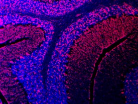
Semaphorin 3E in Mouse Brain.
Semaphorin 3E was detected in immersion fixed frozen sections of adult mouse brain using Goat Anti-Human Semaphorin 3E Antigen Affinity-purified Polyclonal Antibody (Catalog # AF3239) at 10 µg/mL overnight at 4 °C. Tissue was stained using the Northern-Lights™ 557-conjugated Anti-Goat IgG Secondary Antibody (red; Catalog # NL001) and counterstained with DAPI (blue). Specific staining was localized to cerebellum. View our protocol for Fluorescent IHC Staining of Frozen Tissue Sections.Applications for Human/Mouse Semaphorin 3E Antibody
Immunocytochemistry
Sample: Immersion fixed MDA-MB-453 human breast cancer cell line
Immunohistochemistry
Sample: Immersion fixed frozen sections of adult mouse brain
Western Blot
Sample: Recombinant Human Semaphorin 3E (Catalog # 3239-S3)
Formulation, Preparation, and Storage
Purification
Reconstitution
Formulation
Shipping
Stability & Storage
- 12 months from date of receipt, -20 to -70 °C as supplied.
- 1 month, 2 to 8 °C under sterile conditions after reconstitution.
- 6 months, -20 to -70 °C under sterile conditions after reconstitution.
Background: Semaphorin 3E
Semaphorin 3E (Sema3E; previously SemaH) is one of six Class 3 (secreted) semaphorins which in the human share 40-50% amino acid (aa) identity. Class 3 semaphorins are potent chemorepellents that function in axon guidance and/or vascular tip cell guidance during development (1). Sema3E is highly expressed by a subset of motor neurons in developing somites, where it acts as a repulsive cue for PlexinD1-expressing endothelial cells of adjacent intersomitic vessels (2, 3). Crystal structures of semaphorins reveal that the 500 aa N-terminal Sema domain forms a seven-blade b-propeller similar to that found in integrin molecules; 14 conserved cysteine residues and one or more N-glycosylation sites are thought critical for forming the secondary structure (4). C-terminal to the Sema domain, Sema3E has a consensus sequence for furin cleavage which, when used, creates a 61kDa form that does not dimerize and is highly expressed in tumor cell lines with metastatic potential (5, 6). Further C-terminal are a cysteine-knot plexin/semaphorin/integrin (PSI) domain, an Ig-like domain, a cysteine for dimerization and a basic domain containing another furin site. Dimerization and cleavage at the C-terminal site are required for repulsing activity of class 3 semaphorins (7). Human Sema3E shares 90%, 85% and 57% aa identity with mouse, cow and dog Sema3E, respectively. Like other semaphorins, Sema3E signaling is transduced by a transmembrane Plexin dimer, which also has a Sema domain and is coupled to kinase pathways. Unlike other Class 3 semaphorins, Sema3E binds directly to its plexin and does not require interaction with a neuropilin for activity (7). Genetic disruption of either Sema3E or PlexinD1 creates mouse mutants with excessive and disorganized vascular growth and branching, indicating the importance of this ligand-receptor pair for vascular guidance (3, 8).
References
- Eichmann, A. et al. (2005) Genes Dev. 19:1013.
- Cohen, S. et al. (2005) Eur. J. Neurosci. 21:1767.
- Gu, C. et al. (2005) Science 307:265.
- Gherardi, E. et al. (2004) Curr. Opin. Struct. Biol. 14:669.
- Christensen, C. et al. (1998) Cancer Res. 58:1238.
- Christensen, C. et al. (2005) Cancer Res. 65:6167.
- Adams, R. H. et al. (1997) EMBO J. 16:6077.
- Gitler, A. D. et al. (2004) Developmental Cell 7:107.
Alternate Names
Gene Symbol
UniProt
Additional Semaphorin 3E Products
Product Documents for Human/Mouse Semaphorin 3E Antibody
Product Specific Notices for Human/Mouse Semaphorin 3E Antibody
For research use only
