Human Phospho-p70 S6 Kinase (T389) Antibody
R&D Systems, part of Bio-Techne | Catalog # MAB8963
Recombinant Monoclonal Antibody.

Key Product Details
Validated by
Biological Validation
Species Reactivity
Validated:
Human
Cited:
Human, Mouse
Applications
Validated:
Immunocytochemistry, Western Blot
Cited:
Western Blot
Label
Unconjugated
Antibody Source
Recombinant Monoclonal Rabbit IgG Clone # 1045C
Product Specifications
Immunogen
Phosphopeptide containing the human p70 S6 Kinase T389 site
Specificity
Detects human p70 S6 Kinase/p85 S6 Kinase when phosphorylated at T389/T412, respectively.
Clonality
Monoclonal
Host
Rabbit
Isotype
IgG
Scientific Data Images for Human Phospho-p70 S6 Kinase (T389) Antibody
Detection of Human Phospho-p70 S6 Kinase (T389)/p85 S6 Kinase (T412) by Western Blot.
Western blot shows lysates of MCF-7 human breast cancer cell line untreated (-) or treated (+) with 100 ng/mL Recombinant Human IGF-I (Catalog # 291-G1) for 60 minutes. PVDF membrane was probed with 0.1 µg/mL of Rabbit Anti-Human Phospho-p70 S6 Kinase (T389) Monoclonal Antibody (Catalog # MAB8963) followed by HRP-conjugated Anti-Rabbit IgG Secondary Antibody (Catalog # HAF008). Specific bands were detected for Phospho-p70 S6 Kinase (T389) and Phospho-p85 S6 Kinase (T412) at approximately 70 and 85 kDa, respectively (as indicated). This experiment was conducted under reducing conditions and using Immunoblot Buffer Group 1.Phospho-p70 S6 Kinase (T389) in MCF‑7 Human Cell Line.
p70 S6 Kinase phosphorylated at T389 was detected in immersion fixed serum starved MCF-7 human breast cancer cell line untreated (lower panel) or treated with Recombinant Human IGF-I (Catalog # 291-G1) using Rabbit Anti-Human Phospho-p70 S6 Kinase (T389) Monoclonal Antibody (Catalog # MAB8963) at 2 µg/mL for 3 hours at room temperature. Cells were stained using the NorthernLights™ 557-conjugated Anti-Rabbit IgG Secondary Antibody (red; Catalog # NL004) and counterstained with DAPI (blue). Specific staining was localized to cytoplasm and nuclei. View our protocol for Fluorescent ICC Staining of Cells on Coverslips.Detection of Mouse p70 S6 Kinase/S6K by Western Blot
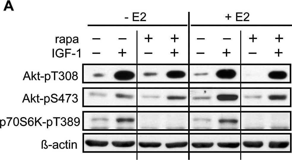
E2 regulates rapamycin effects on mTORC2 activity.A-H, Rapamycin lowers mTORC1 activity independent of presence of E2, ER alpha- or ER beta-agonist, however lowers mTORC2 activity dependent on presence of E2. A-D, HL-1 cells were grown to near confluence in medium containing 10 nM E2, and E-H, 10 nM ER alpha-agonist PPT or 1 nM ER beta-agonist DPN and serum starved for 24 hours prior to incubation with 20 nM rapamycin and IGF-1 for 24 h. A and E show representative westernblots. Equal loading was verified by blotting with antibodies against beta-actin or alpha-tubulin. For B,C,D and F,G,H, labeled bands were quantified with ImageJ software, normalized to loading and IGF-1 induced phosphorylation of indicated proteins was determined by ratio to the value of non-IGF-1 stimulated control cells. Mean ± SEM of fold stimulation by IGF-1 is shown of at least 3 independently performed experiments. * p < 0.05,** p < 0.009, *** p < 0.0001. If not indicated differently, significances are related to respective non-IGF-1 stimulated cells. Image collected and cropped by CiteAb from the following publication (https://dx.plos.org/10.1371/journal.pone.0123385), licensed under a CC-BY license. Not internally tested by R&D Systems.Applications for Human Phospho-p70 S6 Kinase (T389) Antibody
Application
Recommended Usage
Immunocytochemistry
2-25 µg/mL
Sample: Immersion fixed serum starved MCF‑7 human breast cancer cell line untreated or treated with Recombinant Human IGF-I (Catalog # 291-G1)
Sample: Immersion fixed serum starved MCF‑7 human breast cancer cell line untreated or treated with Recombinant Human IGF-I (Catalog # 291-G1)
Western Blot
0.1 µg/mL
Sample: MCF‑7 human breast cancer cell line treated with Recombinant Human IGF‑I (Catalog # 291-G1)
Sample: MCF‑7 human breast cancer cell line treated with Recombinant Human IGF‑I (Catalog # 291-G1)
Formulation, Preparation, and Storage
Purification
Protein A or G purified from cell culture supernatant
Reconstitution
Reconstitute at 0.5 mg/mL in sterile PBS. For liquid material, refer to CoA for concentration.
Formulation
Lyophilized from a 0.2 μm filtered solution in PBS with Trehalose. *Small pack size (SP) is supplied either lyophilized or as a 0.2 µm filtered solution in PBS.
Shipping
Lyophilized product is shipped at ambient temperature. Liquid small pack size (-SP) is shipped with polar packs. Upon receipt, store immediately at the temperature recommended below.
Stability & Storage
Use a manual defrost freezer and avoid repeated freeze-thaw cycles.
- 12 months from date of receipt, -20 to -70 °C as supplied.
- 1 month, 2 to 8 °C under sterile conditions after reconstitution.
- 6 months, -20 to -70 °C under sterile conditions after reconstitution.
Background: p70 S6 Kinase
20-fold in G1 cells released from G0 (2). p70S6K activation requires sequential phosphorylations at proline-directed residues in the putative autoinhibitory pseudosubstrate domain, as well as T389, a site phosphorylated by Phosphoinositide-Dependent Kinase 1 (PDK1).
References
- Ferrari, S. et al. (1994) Crit. Rev. Biochem. Mol. Biol. 29:385.
- Edelmann, H.M. et al. (1996) J. Biol. Chem. 271:963.
Alternate Names
p70-alpha, RPS6KB1, S6K1, STK14A
Gene Symbol
RPS6KB1
Additional p70 S6 Kinase Products
Product Documents for Human Phospho-p70 S6 Kinase (T389) Antibody
Product Specific Notices for Human Phospho-p70 S6 Kinase (T389) Antibody
For research use only
Loading...
Loading...
Loading...
Loading...


