Human Purine Nucleoside Phosphorylase/PNP Antibody
R&D Systems, part of Bio-Techne | Catalog # MAB6486

Key Product Details
Species Reactivity
Validated:
Cited:
Applications
Validated:
Cited:
Label
Antibody Source
Product Specifications
Immunogen
Met1-Ser289
Accession # P00491
Specificity
Clonality
Host
Isotype
Scientific Data Images for Human Purine Nucleoside Phosphorylase/PNP Antibody
Detection of Human Purine Nucleoside Phosphorylase/PNP by Western Blot.
Western blot shows lysates of COLO 205 human colorectal adenocarcinoma cell line, Daudi human Burkitt's lymphoma cell line, HT1080 human fibrosarcoma cell line, and K562 human chronic myelogenous leukemia cell line. PVDF membrane was probed with 0.5 µg/mL of Mouse Anti-Human Purine Nucleoside Phosphorylase/PNP Monoclonal Antibody (Catalog # MAB6486) followed by HRP-conjugated Anti-Mouse IgG Secondary Antibody (Catalog # HAF018). A specific band was detected for Purine Nucleoside Phosphorylase/PNP at approximately 32 kDa (as indicated). This experiment was conducted under reducing conditions and using Immunoblot Buffer Group 1.Purine Nucleoside Phosphorylase/PNP in Human Liver.
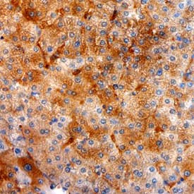
Purine Nucleoside Phosphorylase/PNP was detected in immersion fixed paraffin-embedded sections of human liver using Mouse Anti-Human Purine Nucleoside Phosphorylase/PNP Monoclonal Antibody (Catalog # MAB6486) at 25 µg/mL overnight at 4 °C. Tissue was stained using the Anti-Mouse HRP-DAB Cell & Tissue Staining Kit (brown; Catalog # CTS002) and counter-stained with hematoxylin (blue). Specific staining was localized to endothelial cells in bile canaliculi. View our protocol for Chromogenic IHC Staining of Paraffin-embedded Tissue Sections.Applications for Human Purine Nucleoside Phosphorylase/PNP Antibody
Immunohistochemistry
Sample: Immersion fixed paraffin-embedded sections of human liver
Western Blot
Sample: COLO 205 human colorectal adenocarcinoma cell line, Daudi human Burkitt's lymphoma cell line, HT1080 human fibrosarcoma cell line, and K562 human chronic myelogenous leukemia cell line
Formulation, Preparation, and Storage
Purification
Reconstitution
Formulation
Shipping
Stability & Storage
- 12 months from date of receipt, -20 to -70 °C as supplied.
- 1 month, 2 to 8 °C under sterile conditions after reconstitution.
- 6 months, -20 to -70 °C under sterile conditions after reconstitution.
Background: Purine Nucleoside Phosphorylase/PNP
Purine Nucleoside Phosphorylase (PNP) catalyzes the phophorolysis of N-ribosidic bonds of purine nucleosides and deoxynucleosides. Physiological substrates of PNP include inosine, guanosine, and 2'-deoxyguanosine, but not adenosine (1). PNP is expressed in most tissues, with markedly greater expression in lymphoid tissues. Genetic deficiencies of PNP result in severely compromised T‑lymphocyte function and neurologic dysfunction (2, 3). PNP is used in assays for the measurement of inorganic phosphate (4).
References
- Schramm, V.L. (1998) Annu. Rev. Biochem. 67:693.
- Stoop, W. et al. (1977) N. Eng. J. Med. 296:651.
- Markert, M.L. (1991) Immunodefic. Rev. 3:45.
- Webb, M.R. (1992) Proc. Natl. Acad. Sci. USA. 89:4884.
Alternate Names
Entrez Gene IDs
Gene Symbol
UniProt
Additional Purine Nucleoside Phosphorylase/PNP Products
Product Documents for Human Purine Nucleoside Phosphorylase/PNP Antibody
Product Specific Notices for Human Purine Nucleoside Phosphorylase/PNP Antibody
For research use only

