Human Renin Antibody
R&D Systems, part of Bio-Techne | Catalog # AF4090

Key Product Details
Species Reactivity
Validated:
Cited:
Applications
Validated:
Cited:
Label
Antibody Source
Product Specifications
Immunogen
Leu24-Arg406
Accession # P60016
Specificity
Clonality
Host
Isotype
Scientific Data Images for Human Renin Antibody
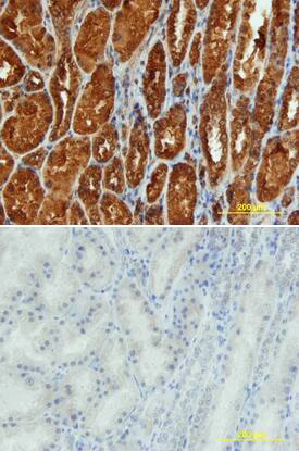
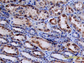
Renin in Human Kidney.
Renin was detected in immersion fixed paraffin-embedded sections of human kidney using Sheep Anti-Human Renin Antigen Affinity-purified Polyclonal Antibody (Catalog # AF4090) at 10 µg/mL overnight at 4 °C. Tissue was stained using the Anti-Sheep HRP-DAB Cell & Tissue Staining Kit (brown; Catalog # CTS019) and counterstained with hematoxylin (blue). View our protocol for Chromogenic IHC Staining of Paraffin-embedded Tissue Sections.Renin in Human Kidney.
Renin was detected in immersion fixed paraffin-embedded sections of human kidney array using Sheep Anti-Human Renin Antigen Affinity-purified Polyclonal Antibody (Catalog # AF4090) at 10 µg/mL overnight at 4 °C. Tissue was stained using the Anti-Sheep HRP-DAB Cell & Tissue Staining Kit (brown; Catalog # CTS019) and counterstained with hematoxylin (blue). Lower panel shows a lack of labeling if primary antibodies are omitted and tissue is stained only with secondary antibody followed by incubation with detection reagents. View our protocol for Chromogenic IHC Staining of Paraffin-embedded Tissue Sections.Applications for Human Renin Antibody
Immunohistochemistry
Sample: Immersion fixed paraffin-embedded sections of human kidney
Immunoprecipitation
Sample: Conditioned cell culture medium spiked with Recombinant Human Renin (Catalog # 4090‑AS), see our available Western blot detection antibodies
Western Blot
Sample: Recombinant Human Renin (Catalog # 4090-AS)
Neutralization
Formulation, Preparation, and Storage
Purification
Reconstitution
Formulation
Shipping
Stability & Storage
- 12 months from date of receipt, -20 to -70 °C as supplied.
- 1 month, 2 to 8 °C under sterile conditions after reconstitution.
- 6 months, -20 to -70 °C under sterile conditions after reconstitution.
Background: Renin
Human Renin is a member of the aspartyl proteinase family produced largely in part by the juxtaglomerular cells in the kidney (1). Renin differs from the other members of this class by having a pH optimum near the neutral pH region with native substrates instead of a pH 2.0 to 3.4 range (2). This more neutral pH optimum allows it to be functional in the plasma. Renin also has a very high selectivity for substrates due to a long peptide recognition on either side of the peptide bond undergoing cleavage. An octapeptide substrate was the minimum length to be cleaved by Renin. Renin plays a crucial role in the regulation of blood pressure and salt balance through the cleavage of angiotensinogen, which is the only known physiological substrate of Renin. Renin releases the decapeptide angiotensin I, which in turn is further converted to vasoactive hormone angiotensin II by angiotensin converting enzyme (ACE). Renin is produced as prorenin with 43 pro residues at the N‑terminal of mature Renin. The inactive prorenin becomes activated proteolytically by trypsin, cathepsin B, or other proteinases.
References
- Yokosawa, H. et al. (1980) J. Biol. Chem. 255:3498.
- Fuminaki, S. et al. (2004) in Handbook of Proteolytic Enzymes, Barret, A. J. et al. eds. p. 54.
Alternate Names
Gene Symbol
UniProt
Additional Renin Products
Product Documents for Human Renin Antibody
Product Specific Notices for Human Renin Antibody
For research use only

