Human RNF4 Antibody
R&D Systems, part of Bio-Techne | Catalog # AF7964

Key Product Details
Validated by
Species Reactivity
Validated:
Cited:
Applications
Validated:
Cited:
Label
Antibody Source
Product Specifications
Immunogen
Met1-Ile190
Accession # P78317
Specificity
Clonality
Host
Isotype
Scientific Data Images for Human RNF4 Antibody
Detection of Human RNF4 by Western Blot.
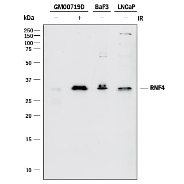
Western blot shows lysates of GM00719D human ataxia telangiectasia cell line either mock-treated (-) or exposed (+) to 10 Gy ionizing radiation (IR) and harvested after 1 hour, BaF3 mouse pro-B cell line, and LNCaP human prostate cancer cell line. PVDF membrane was probed with 1 µg/mL of Goat Anti-Human RNF4 Antigen Affinity-purified Polyclonal Antibody (Catalog # AF7964) followed by HRP-conjugated Anti-Goat IgG Secondary Antibody (Catalog # HAF017). A specific band was detected for RNF4 at approximately 34 kDa (as indicated). This experiment was conducted under reducing conditions and using Immunoblot Buffer Group 1.RNF4 in HeLa Human Cell Line.
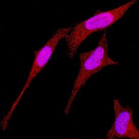
RNF4 was detected in immersion fixed HeLa human cervical epithelial carcinoma cell line using Goat Anti-Human RNF4 Antigen Affinity-purified Polyclonal Antibody (Catalog # AF7964) at 1.7 µg/mL for 3 hours at room temperature. Cells were stained using the NorthernLights™ 557-conjugated Anti-Goat IgG Secondary Antibody (red; Catalog # NL001) and counterstained with DAPI (blue). Specific staining was localized to nuclei and cytoplasm. View our protocol for Fluorescent ICC Staining of Cells on Coverslips.RNF4 in Human Testis.
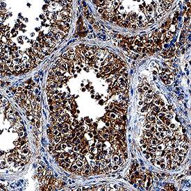
RNF4 was detected in immersion fixed paraffin-embedded sections of human testis using Goat Anti-Human RNF4 Antigen Affinity-purified Polyclonal Antibody (Catalog # AF7964) at 10 µg/mL overnight at 4 °C. Tissue was stained using the Anti-Goat HRP-DAB Cell & Tissue Staining Kit (brown; Catalog # CTS008) and counterstained with hematoxylin (blue). Specific staining was localized to nuclei and cytoplasm. View our protocol for Chromogenic IHC Staining of Paraffin-embedded Tissue Sections.Applications for Human RNF4 Antibody
Immunocytochemistry
Sample: Immersion fixed HeLa human cervical epithelial carcinoma cell line
Immunohistochemistry
Sample: Immersion fixed paraffin-embedded sections of human testis
Western Blot
Sample: GM00719D human ataxia telangiectasia cell line exposed to 10 Gy ionizing radiation (IR) and harvested after 1 hour, BaF3 mouse pro-B cell line, and LNCaP human prostate cancer cell line
Reviewed Applications
Read 1 review rated 4 using AF7964 in the following applications:
Formulation, Preparation, and Storage
Purification
Reconstitution
Formulation
Shipping
Stability & Storage
- 12 months from date of receipt, -20 to -70 °C as supplied.
- 1 month, 2 to 8 °C under sterile conditions after reconstitution.
- 6 months, -20 to -70 °C under sterile conditions after reconstitution.
Background: RNF4
RNF4 (small nuclear ring finger protein, SNURF) is a RING-finger ubiquitin E3 ligase that ubiquitinates and mediates the proteasomal destruction of targets such as PML, PEA3, CENP1, and PARP1. In addition to the RING domain, RNF4 contains four SUMO-interacting motifs (SIMs) that function to recruit this ligase to poly-sumoylated substrates. RNF4 will autoubiquitinate in vitro, and will also ubiquitinate poly-SUMO chains.
References
-
Geoffroy, M-C et al. (2010) Mol. Bio. Cell 21: 4227
-
Tathum, M.H. et al. (2008) Nat. Cell Bio. 10: 538
Long Name
Alternate Names
Entrez Gene IDs
Gene Symbol
UniProt
Additional RNF4 Products
Product Documents for Human RNF4 Antibody
Product Specific Notices for Human RNF4 Antibody
For research use only


