Human S100A13 Antibody
R&D Systems, part of Bio-Techne | Catalog # AF4327

Key Product Details
Species Reactivity
Applications
Label
Antibody Source
Product Specifications
Immunogen
Ala2-Lys98
Accession # Q99584
Specificity
Clonality
Host
Isotype
Scientific Data Images for Human S100A13 Antibody
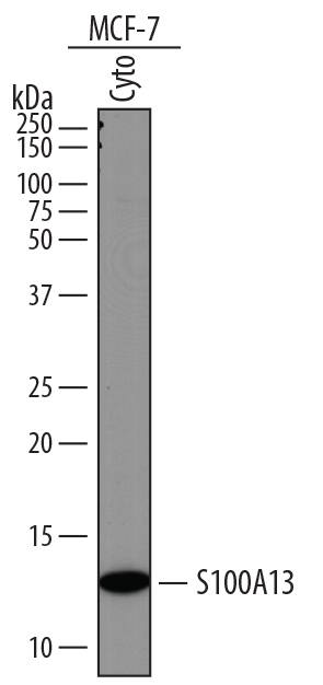
Detection of Human S100A13 by Western Blot.
Western blot shows lysates of MCF-7 human breast cancer cell line. Gels were loaded with 25 µg of cytoplasmic (Cyto) extracts. PVDF membrane was probed with 1 µg/mL of Goat Anti-Human S100A13 Antigen Affinity-purified Polyclonal Antibody (Catalog # AF4327) followed by HRP-conjugated Anti-Goat IgG Secondary Antibody (HAF017). A specific band was detected for S100A13 at approximately 12 kDa (as indicated). This experiment was conducted under reducing conditions and using Immunoblot Buffer Group 1.Applications for Human S100A13 Antibody
Western Blot
Sample: MCF‑7 human breast cancer cell line
Formulation, Preparation, and Storage
Purification
Reconstitution
Formulation
Shipping
Stability & Storage
- 12 months from date of receipt, -20 to -70 °C as supplied.
- 1 month, 2 to 8 °C under sterile conditions after reconstitution.
- 6 months, -20 to -70 °C under sterile conditions after reconstitution.
Background: S100A13
S100A13 is an 11 kDa member of the S100 (soluble in 100% saturated ammonium sulfate) family of vertebrate EF-hand Ca++-binding proteins (1‑3). It is widely expressed as a homodimer with two 98 amino acid (aa) long subunits (2, 3). Human S100A13 shares 83%, 90%, 91%, 87%, 78% and 47% aa identity with mouse, rat, cow, dog, opossum and chicken S100A13, respectively. Like other S100 proteins, S100A13 is small and generally acidic, but contains a basic residue-rich sequence at the C terminus, and two EF hand motifs that bind with Ca++ differing affinities (2‑4). Some S100 proteins, including S100A13, are able to bind the cell surface receptor for advanced glycation end-products (RAGE) (5). Despite lacking a signal sequence, S100A13 plays an important role in Cu++-dependent export of FGF-1 (FGF acidic) and IL-1 alpha from the cell in response to stresses such as heat shock, anoxia and starvation (6‑8). Binding of copper is necessary for formation of a multi-protein complex between S100A13, FGF-1 and p40 synaptotagmin-1 (syt-1) (9, 10). Cu++ ions supplied by S100A13 are thought to oxidize and downregulate the activity of FGF-1 prior to export (10). Calcium influx may also play a similar role in FGF-1 release from neuronal cells (11). S100A13 is composed of four amphiphilic helices that may interact with acidic phospholipid headgroups. With FGF-1 and syt-1, S100A13 likely perturbs the membrane, which allows the S100A13 protein complex to exit the cell (4, 12). S100A13 has been proposed as a marker for angiogenesis in tumors and endometrium, due to its role in stress-induced export of FGF-1 (13, 14). Based on in house studies, S100A13 has also been found to promote neurite outgrowth from rat cortical embryonic neurons (15).
References
- Santamaria-Kisiel, L. et al. (2006) Biochem. J. 396:201.
- Wicki, R. et al. (1996) Biochem. Biophys. Res. Commun. 227:594.
- Ridinger, K. et al. (2000) J. Biol. Chem. 275:8686.
- Li, M. et al. (2007) Biochem. Biophys. Res. Commun. 356:616.
- Hsieh, H.-L. et al. (2004) Biochem. Biophys. Res. Commun. 316:949.
- Landriscina, M. et al. (2001) J. Biol. Chem. 276:22544.
- Sivaraja, V. et al. (2006) Biophys. J. 91:1832.
- Mandinova, A. et al. (2003) J. Cell Sci. 116:2687.
- Prudovsky, I. et al. (2002) J. Cell Biol. 158:201.
- Landriscina, M. et al. (2001) J. Biol. Chem. 276:25549.
- Matsunaga, H. and H. Ueda (2006) Cell. Mol. Neurobiol. 26:237.
- Graziani, I. et al. (2006) Biochem. Biophys. Res. Commun. 349:192.
- Landriscina, M. et al. (2006) J. Neurooncol. 80:251.
- Hayrabedyan, S. et al. (2005) Reprod. Biol. 5:51.
- R&D Sytems (2007) In-house data.
Long Name
Alternate Names
Gene Symbol
UniProt
Additional S100A13 Products
Product Documents for Human S100A13 Antibody
Product Specific Notices for Human S100A13 Antibody
For research use only
