Human Semaphorin 4A Antibody
R&D Systems, part of Bio-Techne | Catalog # MAB46941

Key Product Details
Species Reactivity
Human
Applications
Immunohistochemistry, Western Blot
Label
Unconjugated
Antibody Source
Monoclonal Mouse IgG1 Clone # 741509
Product Specifications
Immunogen
Mouse myeloma cell line NS0-derived recombinant human Semaphorin 4A
Gly32-His683
Accession # Q9H3S1
Gly32-His683
Accession # Q9H3S1
Specificity
Detects human Semaphorin 4A in direct ELISAs and Western blots. In direct ELISA, no cross-reactivity
with recombinant human Semaphorin 4B, 4C, or 4D is observed and approximately
5% cross-reactivity with recombinant mouse Semaphorin 4A is observed.
Clonality
Monoclonal
Host
Mouse
Isotype
IgG1
Scientific Data Images for Human Semaphorin 4A Antibody
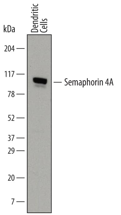
Detection of Human Semaphorin 4A by Western Blot.
Western blot shows lysates of human dendritic cells. PVDF membrane was probed with 2 µg/mL of Mouse Anti-Human Semaphorin 4A Monoclonal Antibody (Catalog # MAB46941) followed by HRP-conjugated Anti-Mouse IgG Secondary Antibody (Catalog # HAF007). A specific band was detected for Semaphorin 4A at approximately 90-95 kDa (as indicated). This experiment was conducted under reducing conditions and using Immunoblot Buffer Group 1.Semaphorin 4A in Mouse Embryo.
Semaphorin 4A was detected in immersion fixed frozen sections of mouse embryo (13 d.p.c.) using Mouse Anti-Human Semaphorin 4A Monoclonal Antibody (Catalog # MAB46941) at 25 µg/mL overnight at 4 °C. Tissue was stained using the Anti-Mouse HRP-DAB Cell & Tissue Staining Kit (brown; Catalog # CTS002) and counterstained with hematoxylin (blue). Specific staining was localized to neuronal processes. View our protocol for Chromogenic IHC Staining of Frozen Tissue Sections.Applications for Human Semaphorin 4A Antibody
Application
Recommended Usage
Immunohistochemistry
8-25 µg/mL
Sample: Immersion fixed frozen sections of mouse embryo (13 d.p.c.)
Sample: Immersion fixed frozen sections of mouse embryo (13 d.p.c.)
Western Blot
2 µg/mL
Sample: Human dendritic cells
Sample: Human dendritic cells
Formulation, Preparation, and Storage
Purification
Protein A or G purified from hybridoma culture supernatant
Reconstitution
Sterile PBS to a final concentration of 0.5 mg/mL. For liquid material, refer to CoA for concentration.
Formulation
Lyophilized from a 0.2 μm filtered solution in PBS with Trehalose. *Small pack size (SP) is supplied either lyophilized or as a 0.2 µm filtered solution in PBS.
Shipping
Lyophilized product is shipped at ambient temperature. Liquid small pack size (-SP) is shipped with polar packs. Upon receipt, store immediately at the temperature recommended below.
Stability & Storage
Use a manual defrost freezer and avoid repeated freeze-thaw cycles.
- 12 months from date of receipt, -20 to -70 °C as supplied.
- 1 month, 2 to 8 °C under sterile conditions after reconstitution.
- 6 months, -20 to -70 °C under sterile conditions after reconstitution.
Background: Semaphorin 4A
References
- Kumanogoh, A. et al. (2003) J. Cell Sci. 116:3463.
- Swissprot Accession # Q9H3S1.
- Entrez Accession # CAI15528, CAI15529, CAI15531, CAI15532, CAI15533 and EAW52993.
- Kumanogoh, A. et al. (2002) Nature 419:629.
- Kumanogoh, A. et al. (2005) Immunity 22:305.
- Yukawa, K. et al. (2005) Int. J. Mol. Med. 16:115.
- Toyofuku, T. et al. (2007) EMBO J. 26:1373.
- Rice, D.S. et al. (2004) Invest. Ophthalmol. Vis. Sci. 45:2767.
- Abid, A. et al. (2007) J. Med. Genet. 43:378.
Alternate Names
CORD10, SEMA4A, SEMAB, SEMB
Gene Symbol
SEMA4A
UniProt
Additional Semaphorin 4A Products
Product Documents for Human Semaphorin 4A Antibody
Product Specific Notices for Human Semaphorin 4A Antibody
For research use only
Loading...
Loading...
Loading...
Loading...

