Human Stanniocalcin 1/STC-1 Antibody
R&D Systems, part of Bio-Techne | Catalog # MAB2958

Key Product Details
Species Reactivity
Validated:
Cited:
Applications
Validated:
Cited:
Label
Antibody Source
Product Specifications
Immunogen
Thr18-Ala247
Accession # P52823
Specificity
Clonality
Host
Isotype
Scientific Data Images for Human Stanniocalcin 1/STC-1 Antibody
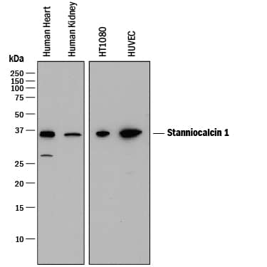
Detection of Human Stanniocalcin 1/STC‑1 by Western Blot.
Western blot shows lysates of human heart tissue, human kidney tissue, HT1080 human fibrosarcoma cell line, and HUVEC human umbilical vein endothelial cells. PVDF membrane was probed with 0.05 µg/mL of Mouse Anti-Human Stanniocalcin 1/STC-1 Monoclonal Antibody (Catalog # MAB2958) followed by HRP-conjugated Anti-Mouse IgG Secondary Antibody (Catalog # HAF018). A specific band was detected for Stanniocalcin 1/STC-1 at approximately 36 kDa (as indicated). This experiment was conducted under reducing conditions and using Immunoblot Buffer Group 1.Detection of Mouse Stanniocalcin 1/STC-1 by Immunocytochemistry/Immunofluorescence
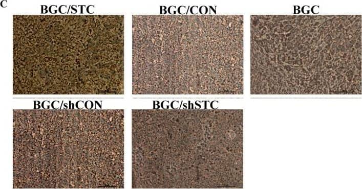
Tumorigenesis and angiogenesis of BGC cells in nude mice. (A) Western blotting analysis of the expression of STC-1 in BGC after stable transfection. (B) Mean volumes of the tumor in each group were calculated. Cultured BGC cells and BGC stable transfection cells (106 cells) were injected subcutaneously into the flank of female nude mice. Tumor volumes were measured and calculated once every two days after we can see the tumor in the flank of nude mice. (C) Immunohistochemical staining of STC-1 in tumor tissues of nude mice. STC-1 was detected on the membrane of tumor cells (D) Immunohistochemical staining of PCNA in tumor tissues of nude mice. PCNA was detected in the nucleus of tumor cells. (E) Quantification of PCNA expression(the Integrated Optical Density (IOD) of PCNA) by image pro-plus software. All histology was carried out on multiple sections from individual mice and three independent in vivo experiments. (F) Hematoxylin and eosin stained sections of the Matrigel plugs (E, endothelial-like cells; T, tumor cells; S, surrounding tissues; V, microvessels). (G) Mean vessel area was quantified in each group. (*P < 0.05, **P < 0.01). Image collected and cropped by CiteAb from the following publication (https://jbiomedsci.biomedcentral.com/articles/10.1186/1423-0127-18-39), licensed under a CC-BY license. Not internally tested by R&D Systems.Applications for Human Stanniocalcin 1/STC-1 Antibody
Western Blot
Sample: Human heart tissue, human kidney tissue, HT1080 human fibrosarcoma cell line, and HUVEC human umbilical vein endothelial cells
Formulation, Preparation, and Storage
Purification
Reconstitution
Formulation
Shipping
Stability & Storage
- 12 months from date of receipt, -20 to -70 °C as supplied.
- 1 month, 2 to 8 °C under sterile conditions after reconstitution.
- 6 months, -20 to -70 °C under sterile conditions after reconstitution.
Background: Stanniocalcin 1/STC-1
Stanniocalcin-1 (STC-1) is a secreted 35 kDa disulfide-linked homodimer that is related to a calcium-regulating hormone in fish. Mature human Stanniocalcin-1 is 230 aa in length, and contains five intrachain disulfide bonds plus one interchain disulfide bond. Circulating Stanniocalcin-1 is both glycosylated and phosphorylated. Mature human Stanniocalcin-1 is 98%, 97%, 95% and 97% aa identical to mature canine, mouse, bovine and rat Stanniocalcin-1, respectively. Stanniocalcin-1 promotes phosphorus absorption and blocks chemokine-induced chemotaxis.
Additional Stanniocalcin 1/STC-1 Products
Product Documents for Human Stanniocalcin 1/STC-1 Antibody
Product Specific Notices for Human Stanniocalcin 1/STC-1 Antibody
For research use only

