Human TBX2 Antibody
R&D Systems, part of Bio-Techne | Catalog # MAB50401

Key Product Details
Species Reactivity
Applications
Label
Antibody Source
Product Specifications
Immunogen
Thr593-Arg702
Accession # Q13207
Specificity
Clonality
Host
Isotype
Scientific Data Images for Human TBX2 Antibody
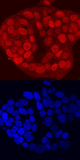
TBX2 in MCF‑7 Human Cell Line.
TBX2 was detected in immersion fixed MCF-7 human breast cancer cell line using Rat Anti-Human TBX2 Monoclonal Antibody (Catalog # MAB50401) at 10 µg/mL for 3 hours at room temperature. Cells were stained using the NorthernLights™ 557-conjugated Anti-Rat IgG Secondary Antibody (red, upper panel; Catalog # NL013) and counterstained with DAPI (blue, lower panel). Specific staining was localized to nuclei. View our protocol for Fluorescent ICC Staining of Cells on Coverslips.Applications for Human TBX2 Antibody
Immunocytochemistry
Sample: Immersion fixed MCF-7 human breast cancer cell line
Formulation, Preparation, and Storage
Purification
Reconstitution
Formulation
Shipping
Stability & Storage
- 12 months from date of receipt, -20 to -70 °C as supplied.
- 1 month, 2 to 8 °C under sterile conditions after reconstitution.
- 6 months, -20 to -70 °C under sterile conditions after reconstitution.
Background: TBX2
T-box protein 2 (TBX2) is a member of the T-box family of transcription factors and functions in the transcriptional regulation of genes required for mesoderm differentiation and limb bud pattern formation. The 74 kDa (predicted MW) nuclear protein, like other T-box family members, contains a T-domain which is involved in DNA binding and dimerization of the protein (aa 99‑277). Human TBX2 shows 96%, 95%, and 71% amino acid identity with dog, mouse, and Xenopus tropicalis TBX2, respectively.
Long Name
Alternate Names
Gene Symbol
UniProt
Additional TBX2 Products
Product Documents for Human TBX2 Antibody
Product Specific Notices for Human TBX2 Antibody
For research use only
