Human Tie-2 APC-conjugated Antibody
R&D Systems, part of Bio-Techne | Catalog # FAB3131A


Key Product Details
Species Reactivity
Validated:
Cited:
Applications
Validated:
Cited:
Label
Antibody Source
Product Specifications
Immunogen
Ala23-Lys745
Accession # AAA61139
Specificity
Clonality
Host
Isotype
Scientific Data Images for Human Tie-2 APC-conjugated Antibody
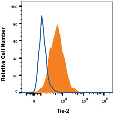
Detection of Tie‑2 in HUVEC Human Cells by Flow Cytometry.
HUVEC human umbilical vein endothelial cells were stained with Mouse Anti-Human Tie-2 APC-conjugated Monoclonal Antibody (Catalog # FAB3131A, filled histogram) or isotype control antibody (Catalog # IC002A, open histogram). View our protocol for Staining Membrane-associated Proteins.Applications for Human Tie-2 APC-conjugated Antibody
Flow Cytometry
Sample: HUVEC human umbilical vein endothelial cells
Formulation, Preparation, and Storage
Purification
Formulation
Shipping
Stability & Storage
- 12 months from date of receipt, 2 to 8 °C as supplied.
Background: Tie-2
Tie-1/Tie (tyrosine kinase with Ig and EGF homology domains 1) and Tie-2/Tek comprise a receptor tyrosine kinase (RTK) subfamily with unique structural characteristics: two immunoglobulin-like domains flanking three epidermal growth factor (EGF)-like domains and followed by three fibronectin type III-like repeats in the extracellular region and a split tyrosine kinase domain in the cytoplasmic region. These receptors are expressed primarily on endothelial and hematopoietic progenitor cells and play critical roles in angiogenesis, vasculogenesis and hematopoiesis.
Human Tie-2 cDNA encodes a 1124 amino acid (aa) residue precursor protein with an 18 residue putative signal peptide, a 727 residue extracellular domain and a 354 residue cytoplasmic domain. Two ligands, angiopoietin-1 (Ang-1) and angiopoietin-2 (Ang-2), which bind Tie-2 with high-affinity have been identified. Ang-2 has been reported to act as an antagonist for Ang-1. Mice engineered to overexpress Ang-2 or to lack Ang-1 or Tie-2 display similar angiogenesis defects.
References
- Partanen, J. and D.J. Dumont (1999) Curr. Top. Microbiol. Immunol. 237:159.
- Takakura, N. et al. (1998) Immunity 9:677.
- Procopio, W. et al. (1999) J. Biol. Chem. 274:30196.
Long Name
Alternate Names
Entrez Gene IDs
Gene Symbol
UniProt
Additional Tie-2 Products
Product Documents for Human Tie-2 APC-conjugated Antibody
Product Specific Notices for Human Tie-2 APC-conjugated Antibody
For research use only