Human TLR3 Antibody
R&D Systems, part of Bio-Techne | Catalog # MAB1487

Key Product Details
Validated by
Species Reactivity
Validated:
Cited:
Applications
Validated:
Cited:
Label
Antibody Source
Product Specifications
Immunogen
Lys27-Ser711
Accession # Q6PCD4
Specificity
Clonality
Host
Isotype
Scientific Data Images for Human TLR3 Antibody
Detection of Human TLR3 by Western Blot.
Western blot shows lysates of Jurkat human acute T cell leukemia cell line and HeLa human cervical epithelial carcinoma cell line. PVDF membrane was probed with 2 µg/mL of Mouse Anti-Human TLR3 Monoclonal Antibody (Catalog # MAB1487) followed by HRP-conjugated Anti-Mouse IgG Secondary Antibody (Catalog # HAF007). A specific band was detected for TLR3 at approximately 116 kDa (as indicated). This experiment was conducted under non-reducing conditions and using Immunoblot Buffer Group 1.Detection of Human TLR3 by Western Blot
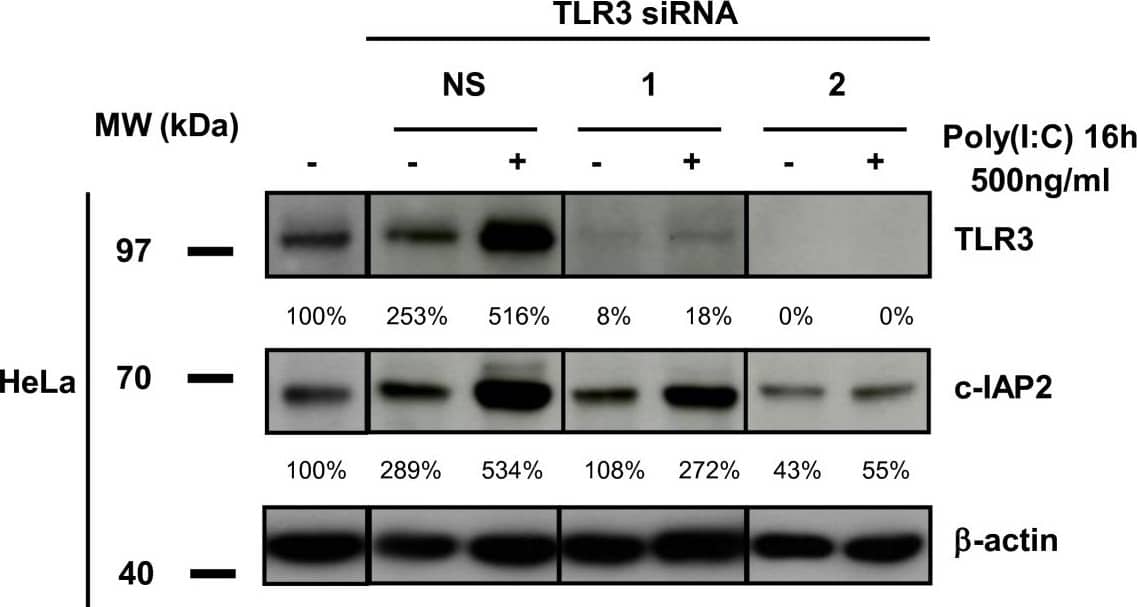
Knocking-down TLR3 suppresses enhancement of c-IAP2 protein expression by poly(I:C). Western blot analysis of protein extracts from HeLa cells treated with poly(I:C) (500 ng/ml for 16 h) with or without pre-treatment with TLR3-specific and control (NS) siRNAs. The same membrane was stained successively with anti-c-IAP2, TLR3 and beta-actin. Note that the control siRNA by itself enhanced TLR3 and c-IAP2 expression. This is consistent with previous observations and possibly related to the interaction of these small double-strand RNAs with the TLR3 itself regardless of their sequence [36]. In our transfection protocol siRNAs are at 100 nM for the first 4 h and 50 nM for the next 44 h. TLR3 inhibition was not complete with the siRNA n°1 which was apparently weaker than the siRNA n°2. However the siRNA n°1 was strong enough to prevent enhancement of TLR3 and c-IAP2 expression in the absence of poly(I:C). Data are representative of two similar experiments. Image collected and cropped by CiteAb from the following publication (https://pubmed.ncbi.nlm.nih.gov/20576118), licensed under a CC-BY license. Not internally tested by R&D Systems.Detection of TLR3 by Western Blot
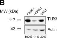
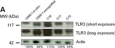
TLR3 is highly expressed in NPC cell lines and xenografts. Western blot analysis reveals intense expression of TLR3 in EBV-positive C666-1, C18, C17 and C15 xenografts/cell lines (A), and in EBV-negative HONE1 and CNE1 NPC cell lines (B). To better characterize the status of TLR3 in NPC cells, its expression was simultaneously assessed in C666-1, in the non NPC head and neck squamous cell carcinoma cell lines SQ20B and FaDu, and in a panel of non malignant and malignant epithelial cell-lines, namely NP69 and NP460 (non tumorigenic immortalized nasopharyngeal cell lines), HeLa, C33 and CaSki (cervical carcinoma), SW613 (colon carcinoma), A549 (non-small cell lung cancer), and A431 (vulvar squamous carcinoma) (C). The same blotted membranes were stained with anti-actin for protein loading controls. Image collected and cropped by CiteAb from the following open publication (https://pubmed.ncbi.nlm.nih.gov/23198710), licensed under a CC-BY license. Not internally tested by R&D Systems.Applications for Human TLR3 Antibody
Western Blot
Sample: Jurkat human acute T cell leukemia cell line and HeLa human cervical epithelial carcinoma cell line
Formulation, Preparation, and Storage
Purification
Reconstitution
Formulation
Shipping
Stability & Storage
- 12 months from date of receipt, -20 to -70 °C as supplied.
- 1 month, 2 to 8 °C under sterile conditions after reconstitution.
- 6 months, -20 to -70 °C under sterile conditions after reconstitution.
Background: TLR3
Human TLR3 is a 116 kDa type I transmembrane glycoprotein that belongs to the mammalian Toll-Like Receptor family of pathogen pattern recognition molecules (1, 2). There are at least eleven mouse and ten human members that activate the innate immune system following exposure to a variety of microbial species (3). The human TLR3 cDNA encodes a 904 amino acid (aa) precursor that contains a 23 aa signal sequence, a 681 aa extracellular domain (ECD), a 21 aa transmembrane segment, and a 179 aa cytoplasmic region (4). The horseshoe shaped ECD (5, 6) contains 23 leucine rich repeats, and the cytoplasmic domain contains one Toll/IL-1 receptor (TIR) domain. The ECD of human TLR3 shares 80%, 79%, and 77% aa sequence identity with the ECD of rat, mouse, and bovine TLR3, respectively. TLR3 is found in phagosomes (7), where the acidic pH enables binding of internalized double stranded RNA and mRNA from viruses, parasites, and necrotic virally-infected cells (8‑11). Ligand binding by TLR3 induces receptor dimerization (5, 6, 8) leading to the release of inflammatory cytokines and dendritic cell maturation (9, 11‑13). TLR3 is expressed in dendritic cells, macrophages, microglia, and astrocytes (13‑15) and is upregulated by IFN-beta and LPS (9, 14). TLR3 expression is also induced by lung fibroblasts and epithelial cells by respiratory syncytial virus infection (12).
References
- Sen, G.C. and S.N. Sarkar (2005) Cytokine Growth Factor Rev. 16:1.
- Schroder, M. and A.G. Bowie (2005) Trends Immunol. 26:462.
- Hopkins, P.A. and S. Sriskandan (2005) Clin. Exp. Immunol. 140:395.
- Rock, F.L. et al. (1998) Proc. Natl. Acad. Sci. 95:588.
- Choe, J. et al. (2005) Science 309:581.
- Bell, J.K. et al. (2005) Proc. Natl. Acad. Sci. 102:10976.
- Nishiya, T. et al. (2002) J. Biol. Chem. 280:37107.
- de Bouteiller, O. et al. (2005) J. Biol. Chem. 280:38133.
- Alexopoulou, L. et al. (2001) Nature 413:732.
- Aksoy, E. et al. (2005) J. Biol. Chem. 280:277.
- Kariko, K. et al. (2004) J. Biol. Chem. 279:12542.
- Rudd, B.D. et al. (2005) J. Virol. 79:3350.
- Farina, C. et al. (2005) J. Neuroimmunol. 159:12.
- Heinz, S. et al. (2003) J. Biol. Chem. 278:21502.
- Wang, T. et al. (2004) Nat. Med. 10:1366.
Long Name
Alternate Names
Gene Symbol
UniProt
Additional TLR3 Products
Product Documents for Human TLR3 Antibody
Product Specific Notices for Human TLR3 Antibody
For research use only


