Human TRIF/TICAM1 Antibody
R&D Systems, part of Bio-Techne | Catalog # MAB6216

Key Product Details
Validated by
Knockout/Knockdown
Species Reactivity
Validated:
Human
Cited:
Human
Applications
Validated:
Western Blot
Cited:
Western Blot
Label
Unconjugated
Antibody Source
Monoclonal Mouse IgG2A Clone # 567212
Product Specifications
Immunogen
E. coli-derived recombinant human TRIF/TICAM1
Met474-Pro618
Accession # Q8IUC6
Met474-Pro618
Accession # Q8IUC6
Specificity
Detects human TRIF/TICAM1 in direct ELISAs and Western blots. In direct ELISAs, no cross-reactivity with recombinant human (rh) TRIF (aa 29‑204), rhTRAM, rhTRIM, rhTRIM5, rhTRIM21, or rhTRIM32 is observed.
Clonality
Monoclonal
Host
Mouse
Isotype
IgG2A
Scientific Data Images for Human TRIF/TICAM1 Antibody
Detection of Human TRIF/TICAM1 by Western Blot.
Western blot shows lysates of Ramos human Burkitt's lymphoma cell line and Raji human Burkitt's lymphoma cell line. PVDF Membrane was probed with 1 µg/mL of Mouse Anti-Human TRIF/TICAM1 Monoclonal Antibody (Catalog # MAB6216) followed by HRP-conjugated Anti-Mouse IgG Secondary Antibody (Catalog # HAF007). A specific band was detected for TRIF/TICAM1 at approximately 113 kDa (as indicated). This experiment was conducted under reducing conditions and using Immunoblot Buffer Group 2.Detection of Human TRIF/TICAM1 by Western Blot
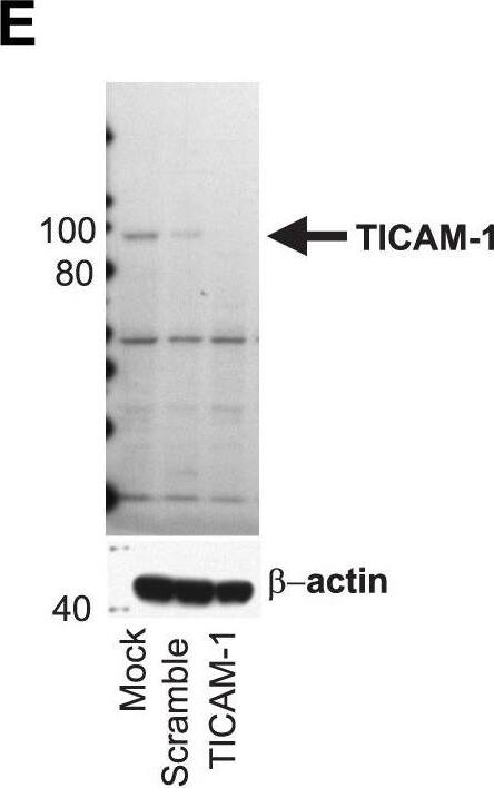
MyD88 is required for inflammasome sensing of HIV and HCV.Knockdowns of the TLR adaptors MyD88 (A, B) and TICAM-1 (TRIF, E, F) were generated and confirmed by previously described RNA interference techniques. (*) denotes comparisons with p≤0.05 compared to the mock transfected cells. Monocytes in which MyD88 was knocked down were cultured with HIVBaL (solid bars) or HCVSubject 180 (hatched bars) and pro-IL-1 beta mRNA transcription measured at 6 h (C, G) and IL-18 secretion measured at 24 h (D, H). Shown are the relative production of pro-IL-1 beta mRNA and IL-18 in MyD88 (C, D) or TICAM-1 (G, H) knockdown monocytes normalized to mock transfected monocytes (no siRNA) stimulated with the same viruses. Bars represent the mean ± S.D. for n = 6–9 independent transfection experiments, (**) denotes comparisons with p≤0.001 compared to the scramble siRNA transfected cells. Image collected and cropped by CiteAb from the following publication (https://dx.plos.org/10.1371/journal.ppat.1004082), licensed under a CC-BY license. Not internally tested by R&D Systems.Detection of Human TRIF/TICAM1 by Western Blot
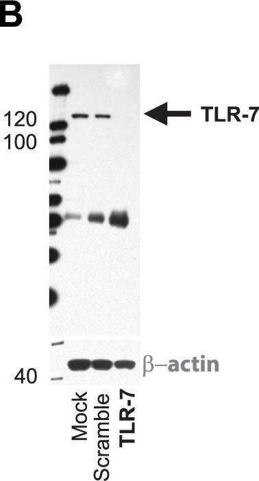
Differential importance of endosomal TLRs in inflammasome sensing of HIV and HCV.Monocytes were cultured without stimulation or with HIVBaL or HCVSubject 180 and IL-1 beta, IFN alpha and measured at multiple timepoints. Fold change in expression of IL-1 beta (black bar) and IFN alpha (grey bar) following HIVBaL or HCVSubject 180 relative to stimulation with media alone is shown at 6 h (A). Functional knockdowns of the endosomal located TLRs in monocytes were generated by RNA interference. Monocytes were mock transfected or transfected with either siRNA targeting TLR7 or a non-targeting sequence (Scramble). After 24 h, cell lysates were prepared and efficacy of knockdown determined by (B) western blot and (C) qRT-PCR. (D) Specificity of the TLR7 siRNA was confirmed by qRT-PCR using primers for TLR3, TLR7, TLR8, and TLR9. (*) denotes comparisons with p≤0.05 compared to the mock transfected cells. Monocytes in which endosomal TLR knockdown were generated were cultured with HIVBaL (solid bars) or HCVSubject 180 (hatched bars) and pro-IL-1 beta mRNA transcription measured at 6 h (E–H) and IL-18 secretion measured at 24 h (I–L). Shown are the relative production of pro-IL-1 beta mRNA and IL-18 in TLR8 (E, I), TLR7 (F, J), TLR3 (G, K) and TLR9 (H, L) knockdown monocytes normalized to mock transfected monocytes (no siRNA) stimulated with the same viruses. Bars represent the mean ± S.D. for n = 6–9 independent transfection experiments, (**) denotes comparisons with p≤0.05 and (***) denoted p≤0.001 compared to the scramble siRNA transfected cells. Image collected and cropped by CiteAb from the following publication (https://dx.plos.org/10.1371/journal.ppat.1004082), licensed under a CC-BY license. Not internally tested by R&D Systems.Applications for Human TRIF/TICAM1 Antibody
Application
Recommended Usage
Western Blot
1 µg/mL
Sample: Ramos human Burkitt's lymphoma cell line and Raji human Burkitt's lymphoma cell line
Sample: Ramos human Burkitt's lymphoma cell line and Raji human Burkitt's lymphoma cell line
Formulation, Preparation, and Storage
Purification
IgM-specific Affinity-purified from hybridoma culture supernatant
Reconstitution
Sterile PBS to a final concentration of 0.5 mg/mL. For liquid material, refer to CoA for concentration.
Formulation
Lyophilized from a 0.2 μm filtered solution in PBS with Trehalose. *Small pack size (SP) is supplied either lyophilized or as a 0.2 µm filtered solution in PBS.
Shipping
Lyophilized product is shipped at ambient temperature. Liquid small pack size (-SP) is shipped with polar packs. Upon receipt, store immediately at the temperature recommended below.
Stability & Storage
Use a manual defrost freezer and avoid repeated freeze-thaw cycles.
- 12 months from date of receipt, -20 to -70 °C as supplied.
- 1 month, 2 to 8 °C under sterile conditions after reconstitution.
- 6 months, -20 to -70 °C under sterile conditions after reconstitution.
Background: TRIF/TICAM1
Long Name
Toll-like receptor adaptor molecule 1
Alternate Names
TICAM1
Gene Symbol
TICAM1
UniProt
Additional TRIF/TICAM1 Products
Product Documents for Human TRIF/TICAM1 Antibody
Product Specific Notices for Human TRIF/TICAM1 Antibody
For research use only
Loading...
Loading...
Loading...
Loading...


