Human Tryptophan Hydroxylase 1/TPH-1 Antibody
R&D Systems, part of Bio-Techne | Catalog # AF5276

Key Product Details
Species Reactivity
Validated:
Cited:
Applications
Validated:
Cited:
Label
Antibody Source
Product Specifications
Immunogen
Met102-Pro403
Accession # P17752
Specificity
Clonality
Host
Isotype
Scientific Data Images for Human Tryptophan Hydroxylase 1/TPH-1 Antibody
Detection of Human Tryptophan Hydroxylase 1/TPH‑1 by Western Blot.
Western blot shows lysates of AGS human gastric adenocarcinoma cell line. PVDF membrane was probed with 1 µg/mL of Goat Anti-Human Tryptophan Hydroxylase 1/TPH-1 Antigen Affinity-purified Polyclonal Antibody (Catalog # AF5276) followed by HRP-conjugated Anti-Goat IgG Secondary Antibody (Catalog # HAF019). A specific band was detected for Tryptophan Hydroxylase 1 at approximately 60 kDa (as indicated). This experiment was conducted under reducing conditions and using Immunoblot Buffer Group 8.Tryptophan Hydroxylase 1/TPH‑1 in Human Brain.
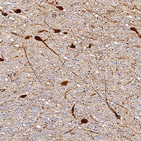
Tryptophan Hydroxylase 1/TPH-1 was detected in immersion fixed paraffin-embedded sections of human brain (substantia nigra) using Goat Anti-Human Tryptophan Hydroxylase 1/TPH-1 Antigen Affinity-purified Polyclonal Antibody (Catalog # AF5276) at 1 µg/mL for 1 hour at room temperature followed by incubation with the Anti-Goat IgG VisUCyte™ HRP Polymer Antibody (Catalog # VC004). Tissue was stained using DAB (brown) and counterstained with hematoxylin (blue). Specific staining was localized to neurons. View our protocol for IHC Staining with VisUCyte HRP Polymer Detection Reagents.Tryptophan Hydroxylase 1/TPH‑1 in Rat Brain.
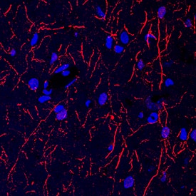
Tryptophan Hydroxylase 1/TPH-1 was detected in perfusion fixed frozen sections of rat brain using Goat Anti-Human Tryptophan Hydroxylase 1/TPH-1 Antigen Affinity-purified Polyclonal Antibody (Catalog # AF5276) at 1.7 µg/mL overnight at 4 °C. Tissue was stained using the NorthernLights™ 557-conjugated Anti-Goat IgG Secondary Antibody (red; Catalog # NL001) and counterstained with DAPI (blue). Specific staining was localized to neurons. View our protocol for Fluorescent IHC Staining of Frozen Tissue Sections.Applications for Human Tryptophan Hydroxylase 1/TPH-1 Antibody
Immunohistochemistry
Sample: Perfusion fixed frozen sections of rat brain and immersion fixed paraffin-embedded sections of human brain (substantia nigra)
Western Blot
Sample: AGS human gastric adenocarcinoma cell line
Formulation, Preparation, and Storage
Purification
Reconstitution
Formulation
Shipping
Stability & Storage
- 12 months from date of receipt, -20 to -70 °C as supplied.
- 1 month, 2 to 8 °C under sterile conditions after reconstitution.
- 6 months, -20 to -70 °C under sterile conditions after reconstitution.
Background: Tryptophan Hydroxylase 1/TPH-1
Tryptophan Hydroxylase 1 (TPH-1) is a 50‑60 kDa intracellular enzyme that belongs to the biopterin-dependent, aromatic amino acid hydroxylase family. It is expressed in gut enterochromaffin cells and pineal gland. TPH-1 catalyzes the hydroxylation of L-Trp, which is the rate-limiting step in serotonin biosynthesis. Human TPH-1 is 444 amino acids (aa) in length. It contains an N-terminal regulatory domain (aa 1‑98), a catalytic region (aa 99‑424), and a C‑terminal Leu-zipper tetramerization domain (aa 425‑444). TPH-1 is phosphorylated on Ser58, and subsequently binds to 14-3-3 proteins, resulting in activation. It functions as a noncovalent Fe-binding 230 kDa homotetramer. There are three potential splice variants. One shows a 29 aa substitution for aa 438‑444, a second shows a 50 aa substitution for aa 157‑444, and a third shows a 68 aa substitution for aa 1‑21. Over aa 102‑403, Human TPH-1 shares 95% aa identical with mouse TPH-1.
Alternate Names
Gene Symbol
UniProt
Additional Tryptophan Hydroxylase 1/TPH-1 Products
Product Documents for Human Tryptophan Hydroxylase 1/TPH-1 Antibody
Product Specific Notices for Human Tryptophan Hydroxylase 1/TPH-1 Antibody
For research use only


