Human VAP-B Antibody
R&D Systems, part of Bio-Techne | Catalog # MAB58551


Key Product Details
Species Reactivity
Validated:
Cited:
Applications
Validated:
Cited:
Label
Antibody Source
Product Specifications
Immunogen
Ala2-Pro132
Accession # O95292
Specificity
Clonality
Host
Isotype
Scientific Data Images for Human VAP-B Antibody
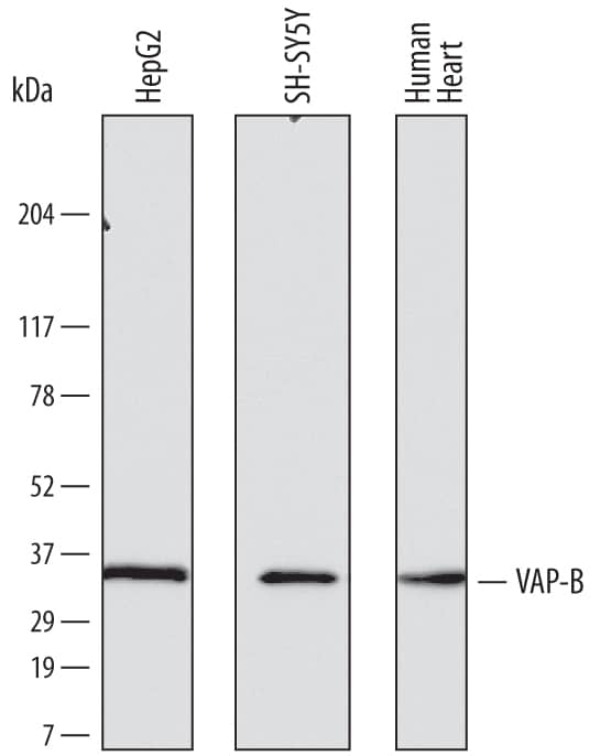
Detection of Human VAP‑B by Western Blot.
Western blot shows lysates of HepG2 human hepatocellular carcinoma cell line, SH-SY5Y human neuroblastoma cell line, and human heart tissue. PVDF membrane was probed with 0.1 µg/mL of Mouse Anti-Human VAP-B Monoclonal Antibody (Catalog # MAB58551) followed by HRP-conjugated Anti-Mouse IgG Secondary Antibody (Catalog # HAF007). A specific band was detected for VAP-B at approximately 32-33 kDa (as indicated). This experiment was conducted under reducing conditions and using Immunoblot Buffer Group 1.VAP‑B in Human Brain.
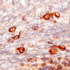
VAP-B was detected in immersion fixed paraffin-embedded sections of human brain (medulla) using Mouse Anti-Human VAP-B Monoclonal Antibody (Catalog # MAB58551) at 15 µg/mL overnight at 4 °C. Before incubation with the primary antibody, tissue was subjected to heat-induced epitope retrieval using Antigen Retrieval Reagent-Basic (Catalog # CTS013). Tissue was stained using the Anti-Mouse HRP-DAB Cell & Tissue Staining Kit (brown; Catalog # CTS002) and counterstained with hematoxylin (blue). Specific staining was localized to the cytoplasm of neurons. View our protocol for Chromogenic IHC Staining of Paraffin-embedded Tissue Sections.Detection of VAPB in T98G Human Cell Line by Flow Cytometry.
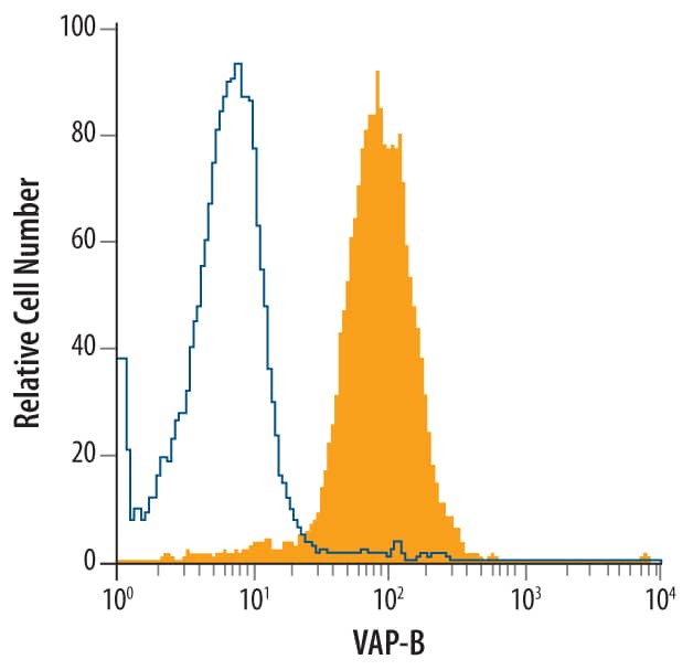
T98G human glioblastoma cell line was stained with Mouse Anti-Human VAP-B Monoclonal Antibody (Catalog # MAB58551, filled histogram) or isotype control antibody (Catalog # MAB002, open histogram), followed by Allophycocyanin-conjugated Anti-Mouse IgG Secondary Antibody (Catalog # F0101B). To facilitate intracellular staining, cells were fixed with paraformaldehye and permeabilized with saponin.Applications for Human VAP-B Antibody
CyTOF-ready
Immunohistochemistry
Sample: Immersion fixed paraffin-embedded sections of human brain (medulla)
Intracellular Staining by Flow Cytometry
Sample: T98G human glioblastoma cell line fixed with paraformaldehye and permeabilized with saponin.
Western Blot
Sample: HepG2 human hepatocellular carcinoma cell line, SH‑SY5Y human neuroblastoma cell line, and human heart tissue
Reviewed Applications
Read 1 review rated 5 using MAB58551 in the following applications:
Formulation, Preparation, and Storage
Purification
Reconstitution
Formulation
Shipping
Stability & Storage
- 12 months from date of receipt, -20 to -70 °C as supplied.
- 1 month, 2 to 8 °C under sterile conditions after reconstitution.
- 6 months, -20 to -70 °C under sterile conditions after reconstitution.
Background: VAP-B
Vesicle-associated membrane protein (VAMP)-associated protein B (VAP-B; also VAMP-B) is an ~30 kDa ubiquitously expressed type IV transmembrane protein belonging to the VAP family (1, 2). It is found in endoplasmic reticulum (ER), Golgi and other membranes as a homodimer or a heterodimer with VAP-A, probably associating through a GxxxG motif in the transmembrane regions (1, 2). Human VAP-B cDNA encodes 243 amino acids (aa) that include a 222 aa cytoplasmic domain and a 21 aa C-terminal membrane anchor. The cytoplasmic domain contains a mobile sperm protein (MSP) domain (aa 7‑124) and a coiled-coil region (aa 159‑196). Human VAP-B shares 90%, 89%, 96%, 96% and 94% aa identity with mouse, rat, canine, bovine and porcine VAP-B, respectively. VAP-A and VAP-B MSP domains recruit FFAT (two phenylalanines in an acidic tract)-motif-containing proteins to the cytosolic surface of ER membranes (2‑4). FFAT proteins mediate many of the effects of VAPs on regulation of membrane transport, phospholipid biosynthesis, microtubule organization, and the unfolded protein response (2, 3). VAPs also interact with some SNARE and viral proteins (2). A human polymorphism of VAP-B, P56S, is found in three familial motor neuron diseases, notably the amylotrophic lateral sclerosis variant ALS8 (2). It produces a non-functional protein that can dimerize with and inhibit function of normal VAP-B, leading formation of intracellular aggregates and increased ER-stress-induced death of motor neurons (5‑7). It can also promote cleavage and secretion of soluble VAP-B, which can then function as a ligand for EPH receptors (8). A naturally occurring 99 aa isoform of VAP-B that diverges at aa 71 within the MSP domain is termed VAP-C (1, 9). It also appears to be a negative regulator of VAP-A and VAP-B (9). While VAP-B is used by hepatitis C virus (HCV) for its propagation, VAP-C inhibits HCV propagation (9).
References
- Nishimura, Y. et al. (1999) Biochem. Biophys. Res. Commun. 254:21.
- Lev, S. et al. (2008) Trends Cell Biol. 18:282.
- Peretti, D. et al., 2008, Mol. Biol. Cell 19:3871.
- Kaiser, S.E. et al., 2005, Structure 13:1035.
- Prosser, D.C. et al. (2008) J. Cell Sci. 121:3052.
- Gkogkas, C. et al. (2008) Hum. Mol. Genet. 17:1517.
- Suzuki, H. et al. (2009) J. Neurochem. 108:973.
- Tsuda, H. et al. (2008) Cell 133:963.
- Kukihara, H. et al. (2009) J. Virol. 83:7959.
Long Name
Alternate Names
Gene Symbol
UniProt
Additional VAP-B Products
Product Documents for Human VAP-B Antibody
Product Specific Notices for Human VAP-B Antibody
For research use only

