Human WARP Antibody
R&D Systems, part of Bio-Techne | Catalog # MAB6189

Key Product Details
Species Reactivity
Applications
Label
Antibody Source
Product Specifications
Immunogen
Arg19-Pro445
Accession # Q6PCB0
Specificity
Clonality
Host
Isotype
Scientific Data Images for Human WARP Antibody
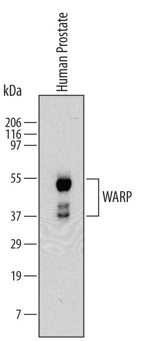
Detection of Human WARP by Western Blot.
Western blot shows lysates of human prostate tissue. PVDF Membrane was probed with 2 µg/mL of Mouse Anti-Human WARP Monoclonal Antibody (Catalog # MAB6189) followed by HRP-conjugated Anti-Mouse IgG Secondary Antibody (Catalog # HAF007). Specific bands were detected for WARP at approximately 50, 40 and 37 kDa (as indicated). This experiment was conducted under reducing conditions and using Immunoblot Buffer Group 1.Applications for Human WARP Antibody
Western Blot
Sample: Human prostate tissue
Formulation, Preparation, and Storage
Purification
Reconstitution
Formulation
Shipping
Stability & Storage
- 12 months from date of receipt, -20 to -70 °C as supplied.
- 1 month, 2 to 8 °C under sterile conditions after reconstitution.
- 6 months, -20 to -70 °C under sterile conditions after reconstitution.
Background: WARP
Von Willebrand factor A (vWFA) domain-related protein (WARP) is a 50 kDa glycoprotein member of the vWFA domain superfamily of extracellular matrix proteins. It is expressed in embryonic articular cartilage, skeletal muscle and basement membranes in the PNS. WARP forms disulfide-linked homodimers and multimers, and complexes with perlecan. Secreted human WARP contains a vWFA domain (aa 34‑213), two fibronectin type III domains (aa 211‑301 and 331‑421) that likely bind to the GAG modification of perlecan, and one potential site for N-linked glycosylation. There is one alternate start site at Met213. Mature human WARP shares 72% aa sequence identity with mature mouse and rat WARP.
Long Name
Alternate Names
Gene Symbol
UniProt
Additional WARP Products
Product Documents for Human WARP Antibody
Product Specific Notices for Human WARP Antibody
For research use only
