Mouse CD40 Ligand/TNFSF5 Antibody
R&D Systems, part of Bio-Techne | Catalog # MAB1163


Key Product Details
Species Reactivity
Validated:
Cited:
Applications
Validated:
Cited:
Label
Antibody Source
Product Specifications
Immunogen
Glu61-Leu260
Accession # P27548
Specificity
Clonality
Host
Isotype
Endotoxin Level
Scientific Data Images for Mouse CD40 Ligand/TNFSF5 Antibody
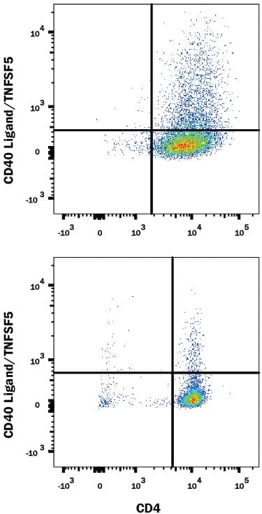
Detection of CD40 Ligand/TNFSF5 in Mouse Splenocytes by Flow Cytometry.
Mouse splenocytes either (A) stimulated to induce Th1 cells or (B) unstimulated were stained with Rat Anti-Mouse CD40 Ligand/TNFSF5 Monoclonal Antibody (Catalog # MAB1163) followed by Allophycocyanin-conjugated Anti-Rat IgG Secondary Antibody (Catalog # F0113) and Rat Anti-Mouse CD4 PE-conjugated Monoclonal Antibody (Catalog # FAB554P). Quadrant markers were set based on control antibody staining (Catalog # MAB006). View our protocol for Staining Membrane-associated Proteins.Proliferation Induced by CD40 Ligand/TNFSF5 and Neutralization by Mouse CD40 Ligand/TNFSF5 Antibody.
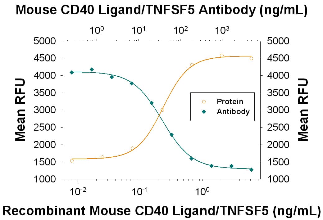
Recombinant Mouse CD40 Ligand/TNFSF5 (Catalog # 8230-CL) induces proliferation in mouse splenic B cells in a dose-dependent manner (orange line), as measured by Resazurin (Catalog # AR002). Proliferation elicited by Recombinant Mouse CD40 Ligand/TNFSF5 (0.5 ng/mL) is neutralized (green line) by increasing concentrations of Rat Anti-Mouse CD40 Ligand/TNFSF5 Monoclonal Antibody (Catalog # MAB1163). The ND50 is typically 0.0075-0.045 µg/mL.Applications for Mouse CD40 Ligand/TNFSF5 Antibody
CyTOF-ready
Flow Cytometry
Sample: Mouse splenocytes stimulated to induce Th1 cells
Neutralization
Formulation, Preparation, and Storage
Purification
Reconstitution
Formulation
Shipping
Stability & Storage
- 12 months from date of receipt, -20 to -70 °C as supplied.
- 1 month, 2 to 8 °C under sterile conditions after reconstitution.
- 6 months, -20 to -70 °C under sterile conditions after reconstitution.
Background: CD40 Ligand/TNFSF5
CD40 ligand (CD40L; also known as CD154, TNFSF5, TRAP, or gp39) is a 260 amino acid (aa) type II transmembrane glycoprotein belonging to the TNF family. Mouse CD40L consists of a 22 aa cytoplasmic domain, a 24 aa transmembrane domain, and 214 aa extracellular domain bearing a single glycosylation site (1, 2). CD40L is expressed predominantly on activated CD4+ T lymphocytes and also found in other types of cells, including NK cells, mast cells, basophils, and eosinophils. Murine CD40L shares 78% amino acid sequence identity with human CD40L. Native bioactive soluble CD40L exists. Soluble human trimeric CD40L secreted by stimulated T cells has been shown to be generated by proteolysis in the microsomes (3). Both membrane bound and soluble CD40L induce similar effects on B cells (3, 4). The receptor of CD40L is CD40, a type I transmembrane glycoprotein belonging to the TNF receptor family. CD40 is expressed on B lymphocytes, monocytes, dendritic cells, and thymic epithelium. Although all monomeric, dimeric, and trimeric forms of soluble CD40L can bind to CD40, the soluble trimeric form of CD40L has the most potent biological activity through oligomerization of cell surface CD40, a common feature of TNF receptor family members (2). The genetic defect in the hyper-IgM syndrome is due to point mutations or deletions of the gene encoding the CD40L, which prevent CD40L from interacting with CD40 (5‑7). CD40L mediates a range of activities on B cells including induction of activation-associated surface antigen, entry into the cell cycle, isotype switching, Ig secretion, and memory generation (8, 9). CD40-CD40L interaction also plays important roles in monocyte activation and dendritic cell maturation (10).
References
- Armitage, R.J. et al. (1992) Nature 357:80.
- Hollenbaugh, D. et al. (1992) EMBO J. 11:4313.
- Fabienne, P. et al. (1996) J. Biol. Chem. 271:5965.
- Fabienne, P. et al. (1996) Eur. J. Immunol 26:725.
- Arrufo, A. et al. (1993) Cell 72:291.
- Hill, A. and N. Chapel et al. (1993) Nature 361:494.
- Korthauer, U. et al. (1993) Nature 361:539.
- Spriggs, M.K. et al. (1992) J. Exp. Med. 176:1543.
- Fanslow, W.C. et al. (1994) Seminars in Immunology 6:267.
- Kooten, C.V. and J. Banchereau (2000) J. Leukoc. Biol. 67:2.
Alternate Names
Gene Symbol
UniProt
Additional CD40 Ligand/TNFSF5 Products
- All Products for CD40 Ligand/TNFSF5
- CD40 Ligand/TNFSF5 cDNA Clones
- CD40 Ligand/TNFSF5 ELISA Kits
- CD40 Ligand/TNFSF5 Luminex Assays
- CD40 Ligand/TNFSF5 Lysates
- CD40 Ligand/TNFSF5 Primary Antibodies
- CD40 Ligand/TNFSF5 Proteins and Enzymes
- CD40 Ligand/TNFSF5 Simple Plex
- CD40 Ligand/TNFSF5 Small Molecules and Peptides
Product Documents for Mouse CD40 Ligand/TNFSF5 Antibody
Product Specific Notices for Mouse CD40 Ligand/TNFSF5 Antibody
For research use only
