Mouse CXADR Antibody
R&D Systems, part of Bio-Techne | Catalog # AF2654


Key Product Details
Species Reactivity
Validated:
Cited:
Applications
Validated:
Cited:
Label
Antibody Source
Product Specifications
Immunogen
Leu20-Gly237
Accession # P97792
Specificity
Clonality
Host
Isotype
Scientific Data Images for Mouse CXADR Antibody
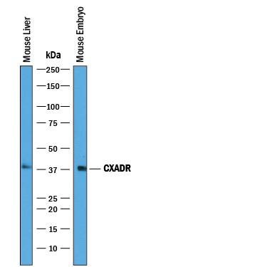
Detection of Mouse CXADR by Western Blot.
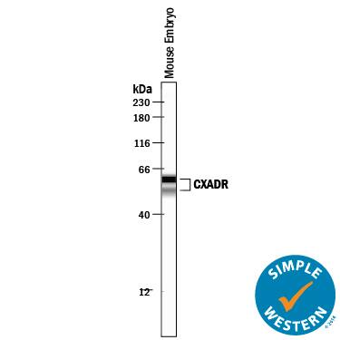
Western blot shows lysates of mouse liver tissue and mouse embryo tissue. PVDF membrane was probed with 0.25 µg/mL of Goat Anti-Mouse CXADR Antigen Affinity-purified Polyclonal Antibody (Catalog # AF2654) followed by HRP-conjugated Anti-Goat IgG Secondary Antibody (Catalog # HAF019). A specific band was detected for CXADR at approximately 40 kDa (as indicated). This experiment was conducted under reducing conditions and using Immunoblot Buffer Group 1.Detection of Mouse CXADR by Simple WesternTM.
Simple Western lane view shows lysates of mouse embryo tissue, loaded at 0.2 mg/mL. Specific bands were detected for CXADR at approximately 60 and 53 kDa (as indicated) using 12.5 µg/mL of Goat Anti-Mouse CXADR Antigen Affinity-purified Polyclonal Antibody (Catalog # AF2654) followed by 1:50 dilution of HRP-conjugated Anti-Goat IgG Secondary Antibody (Catalog # HAF109). This experiment was conducted under reducing conditions and using the 12-230 kDa separation system.Applications for Mouse CXADR Antibody
Immunohistochemistry
Sample: Immersion fixed frozen sections of mouse embryo (E15.5)
Simple Western
Sample: Mouse embryo tissue
Western Blot
Sample: Mouse liver tissue and mouse embryo tissue
Formulation, Preparation, and Storage
Purification
Reconstitution
Formulation
Shipping
Stability & Storage
- 12 months from date of receipt, -20 to -70 °C as supplied.
- 1 month, 2 to 8 °C under sterile conditions after reconstitution.
- 6 months, -20 to -70 °C under sterile conditions after reconstitution.
Background: CXADR
CXADR (Coxsackie and Adenovirus Receptor), also known as CAR, is a 46 kDa type I transmembrane glycoprotein that belongs to the CTX family of the Ig superfamily (1‑3). CXADR has received attention as a receptor that facilitates gene transfer mediated by most adenoviruses (1, 2). It is also an adhesion molecule within junctional complexes, notably between epithelial cells lining body cavities and within myocardial intercalated discs (1, 2, 4). CXADR is essential for normal cardiac development in the mouse (7). It is expressed throughout brain neuroepithelium during development, but mainly in ependymal cells in the adult (4‑6). The 365 amino acid (aa) mouse CXADR contains a 19 aa signal sequence, a 218 aa extracellular domain (ECD) with a V-type (D1) and a C2-type (D2) Ig-like domain, a 21 aa transmembrane segment and a 107 aa intracellular domain. D1 is thought to be responsible for homodimer formation in trans within tight junctions (2). The fiber knob of adenoviruses attaches at a similar site, and evidence suggests that disruption of tight junctions facilitates virus binding (1, 2). A PDZ binding motif at the C‑terminus interacts with several cytoplasmic junctional proteins (1). The ECD of mouse CXADR shares 97%, 90%, 89%, 89% and 88% aa sequence identity with the corresponding regions of rat, human, bovine, porcine and canine CXADR, respectively. An alternately spliced isoform (CXADR2) that diverges in the C‑terminal 15 aa shows the same expression pattern, but may show different subcellular localization (4, 8). Transcription of other splice variants has been detected, but not their translation. A secreted form identified in serum and pleural fluid can block viral infection (9).
References
- Coyne, C.B. and J.M. Bergelson (2005) Adv. Drug Deliv. Rev. 57:869.
- Philipson, L. and R.F. Pettersson (2004) Curr. Top. Microbiol. Immunol. 273:87.
- Tomko, R.P. et al. (1997) Proc. Natl. Acad. Sci. USA 94:3352.
- Raschperger, E. et al. (2006) Exp. Cell Res. 312:1566.
- Hotta, Y. et al. (2003) Dev. Brain Res. 143:1.
- Hauwel, M. et al. (2005) Brain Res. Rev. 48:265.
- Chen, J. et al. (2006) Circ. Res. 98:923.
- Mirza, M. et al. (2006) Exp. Cell Res. 312:817.
- Bernal, R.M. et al. (2002) Clin. Cancer Res. 8:1915.
Long Name
Alternate Names
Gene Symbol
UniProt
Additional CXADR Products
Product Documents for Mouse CXADR Antibody
Product Specific Notices for Mouse CXADR Antibody
For research use only
