Mouse DMP-1 Antibody
R&D Systems, part of Bio-Techne | Catalog # AF4386

Key Product Details
Species Reactivity
Validated:
Cited:
Applications
Validated:
Cited:
Label
Antibody Source
Product Specifications
Immunogen
Leu17-Tyr503
Accession # Q2HJ09
Specificity
Clonality
Host
Isotype
Scientific Data Images for Mouse DMP-1 Antibody
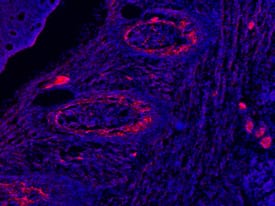
DMP-1 in Embryonic Mouse Rib.
DMP-1 was detected in immersion fixed frozen sections of embryonic mouse rib (E15.5) using 10 µg/mL Sheep Anti-Mouse DMP-1 Antigen Affinity-purified Polyclonal Antibody (Catalog # AF4386) overnight at 4 °C. Tissue was stained with the NorthernLights™ 557-conjugated Anti-Sheep IgG Secondary Antibody (red; NL010) and counterstained with DAPI (blue). View our protocol for Fluorescent IHC Staining of Frozen Tissue Sections.Detection of Mouse DMP-1 by Immunohistochemistry
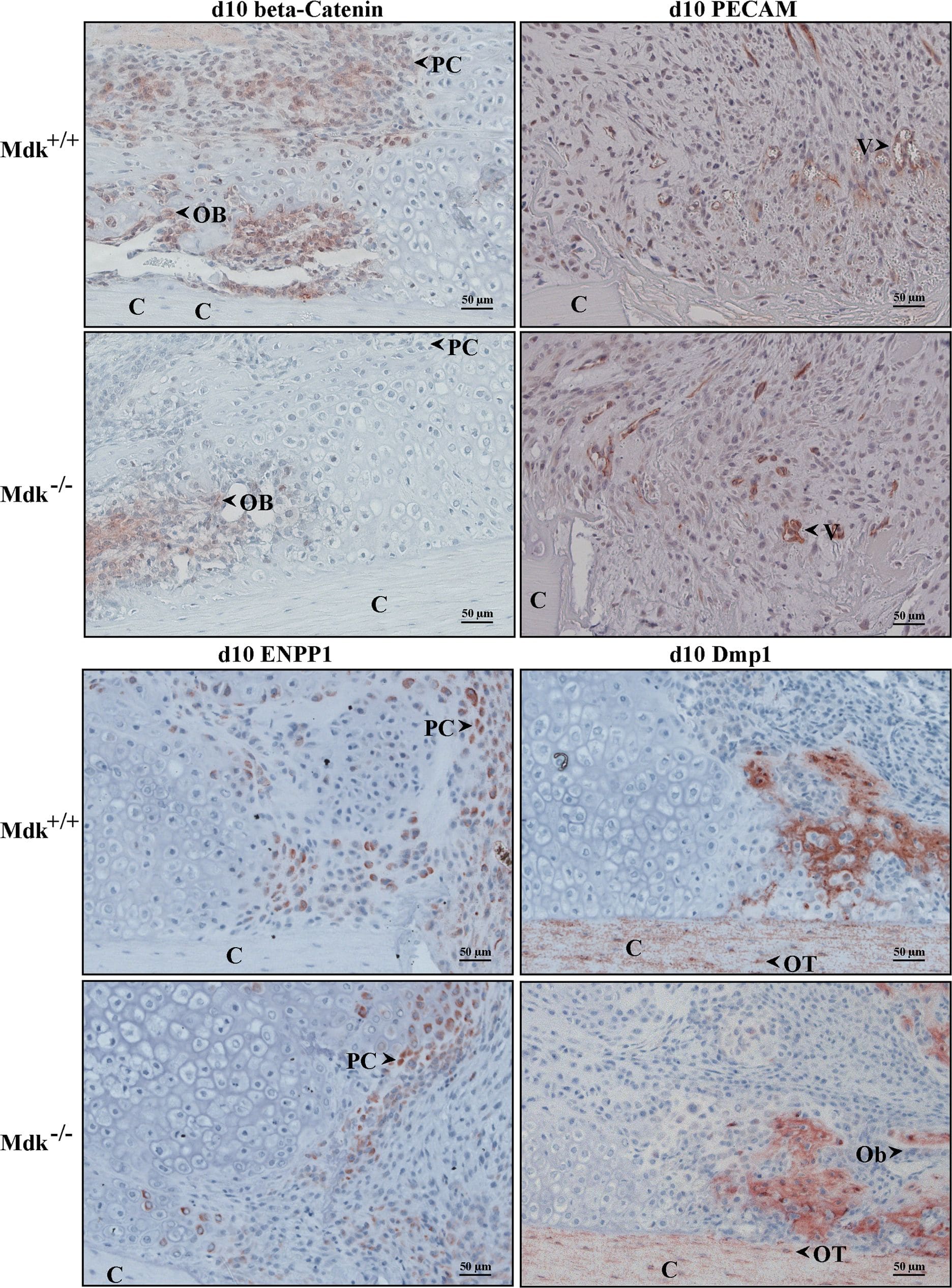
Immunohistochemical staining showing reduced beta-catenin levels in chondrocytes of Mdk-deficient mice.Sections of fractured femurs from four mice of each time point and genotype group were stained for each antigen and counterstained using hematoxylin. Representative images are shown; C = cortex; PC = proliferating chondrocyte; Ob = osteoblast; V = vessel; OT = osteocyte; scale bar 50 µm; 200-fold magnification. Beta-catenin staining of the periosteal callus at day 10. PECAM staining of the periosteal fracture callus bridging the osteotomy gap showing the endothelial cells of the newly formed vessels in an area of proliferating chondrocytes at day 10. Enpp1 staining of the osteotomy gap at day 10 showing positively stained proliferating chondrocytes. Dmp1 staining of the periosteal callus at day 10 showing positively stained cortex, osteocytes and areas of new bone formation. Osteoblasts were Dmp1 negative. (n = 4 per group). Image collected and cropped by CiteAb from the following publication (https://pubmed.ncbi.nlm.nih.gov/25551381), licensed under a CC-BY license. Not internally tested by R&D Systems.Applications for Mouse DMP-1 Antibody
Immunohistochemistry
Sample: Immersion fixed frozen sections of embryonic mouse rib (E15.5)
Western Blot
Sample: Recombinant Mouse DMP-1 (Catalog # 4386-DM)
Formulation, Preparation, and Storage
Purification
Reconstitution
Formulation
Shipping
Stability & Storage
- 12 months from date of receipt, -20 to -70 °C as supplied.
- 1 month, 2 to 8 °C under sterile conditions after reconstitution.
- 6 months, -20 to -70 °C under sterile conditions after reconstitution.
Background: DMP-1
Dentin matrix protein 1 (DMP-1) is a member of the SIBLING family of proteins that includes bone sialoprotein, dentin sialophosphoprotein, MEPE, and osteopontin. These are highly phosphorylated integrin-binding proteins that are rich in acidic amino acids and function in the formation of calcified bone and tooth matrix (1, 2). Its phosphate content, spacing of acidic residues, and calcium-dependent dimerization of DMP-1 contribute to its ability to sequester calcium phosphate clusters and promote hydroxyapatite (HA) crystal formation (3-5). Mature mouse DMP-1 is 487 amino acids (aa) in length. It contains a poly-Pro segment (aa 41-44) and an RGD binding motif (aa 350-352). DMP-1 may be cleaved by BMP-1 family proteases at a single site which is conserved in human, generating a 37 kDa N-terminal (aa 17‑212) and a 57 kDa C-terminal (aa 213-503) fragment (6). The N-terminal fragment in rat carries chondroitin sulfate (7). The C-terminal fragment alone can nucleate HA crystals, while crystal growth into a needle-like morphology is inhibited by the N-terminal fragment (3, 4). Crystal maturation is dependent on the presence of type I collagen (4). DMP-1 is required for odontoblast differentiation as well as dentin formation (8). Unphosphorylated DMP-1 is retained intracellularly where it is targeted to the nucleus. Here, it activates the transcription of odontoblast and osteoblast specific genes (9, 10). Early in osteoblast maturation, nuclear DMP-1 is extensively phosphorylated by casein kinase II, triggering its secretion (9). DMP-1 mutations in humans are associated with hypophosphatemia and FGF-23 over-expression (11, 12). DMP-1 induces the activation of pro-MMP-9 and displaces mature MMP-9 from TIMP1 (13). DMP-1 tethers MMP-9 to the cell surface via CD44 and integrins alphaV beta3 and alphaV beta5, promoting tumor cell invasiveness in vitro (14). Full length DMP-1 circulates in human serum in a tight complex with complement factor H (13, 14). When first bound to CD44 or integrin alphaV beta3, DMP-1 can anchor factor H to the cell surface and protect the cell from complement-mediated lysis (15). Mature mouse DMP-1 shares 63%, 61%, and 87% aa sequence identity with bovine, human, and rat DMP-1, respectively.
References
- Qin, C. et al. (2004) Crit. Rev. Oral Biol. Med. 15:126.
- MacDougall, M. et al. (1998) J. Bone Miner. Res. 13:422.
- He, G. et al. (2003) Nat. Mater. 2:552.
- Gajjeraman, S. et al. (2007) J. Biol. Chem. 282:1193.
- He, G. et al. (2005) Biochemistry 44:16140.
- Steiglitz, B.M. et al. (2004) J. Biol. Chem. 279:980.
- Qin, C. et al. (2006) J. Biol. Chem. 281:8034.
- Lu, Y. et al. (2007) Dev. Biol. 303:191.
- Narayanan, K. et al. (2003) J. Biol. Chem. 278:17500.
- Narayanan, K. et al. (2006) J. Biol. Chem. 281:19064.
- Lorenz-Depiereux, B. et al. (2006) Nat. Genet. 38:1248.
- Feng, J.Q. et al. (2006) Nat. Genet. 38:1310.
- Fedarko, N.S. et al. (2004) FASEB J. 18:735.
- Karadag, A. et al. (2005) Cancer Res. 65:11545.
- Jain, A. et al. (2002) J. Biol. Chem. 277:13700.
Long Name
Alternate Names
Gene Symbol
UniProt
Additional DMP-1 Products
Product Documents for Mouse DMP-1 Antibody
Product Specific Notices for Mouse DMP-1 Antibody
For research use only

