Mouse ECM1 Antibody
R&D Systems, part of Bio-Techne | Catalog # AF4428

Key Product Details
Species Reactivity
Applications
Label
Antibody Source
Product Specifications
Immunogen
Ala20-Glu559
Accession # AAI38694
Specificity
Clonality
Host
Isotype
Scientific Data Images for Mouse ECM1 Antibody
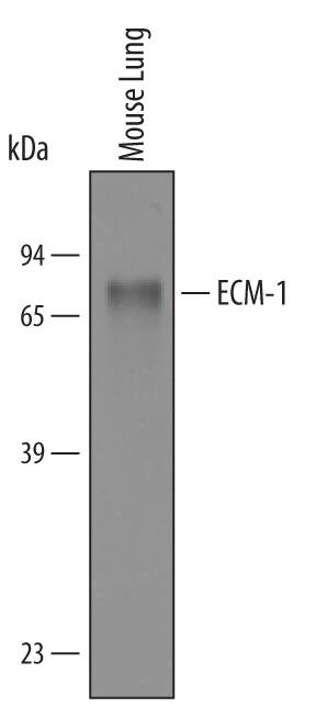
Detection of Mouse ECM-1 by Western Blot.
Western blot shows lysates of mouse lung tissue. PVDF membrane was probed with 1 µg/mL of Goat Anti-Mouse ECM-1 Antigen Affinity-purified Polyclonal Antibody (Catalog # AF4428) followed by HRP-conjugated Anti-Goat IgG Secondary Antibody (Catalog # HAF019). A specific band was detected for ECM-1 at approximately 85 kDa (as indicated). This experiment was conducted under reducing conditions and using Immunoblot Buffer Group 8.Applications for Mouse ECM1 Antibody
Western Blot
Sample: Mouse lung tissue
Formulation, Preparation, and Storage
Purification
Reconstitution
Formulation
Shipping
Stability & Storage
- 12 months from date of receipt, -20 to -70 °C as supplied.
- 1 month, 2 to 8 °C under sterile conditions after reconstitution.
- 6 months, -20 to -70 °C under sterile conditions after reconstitution.
Background: ECM1
Extracellular matrix protein-1 (ECM-1, ECM-1a) is an 85 kDa, secreted glycoprotein important in connective tissue organization (1‑3). Of identified splice variants, the 559 amino acid (aa) form, ECM-1a is most widely expressed, with highest expression in the placenta, heart, and developing bones (3, 4). ECM-1b (434 aa) is found only in tonsil and skin, where it is associated with suprabasal keratinocytes (3, 5). Mouse ECM-1 contains a 19 aa signal peptide and a 540 aa secreted portion that includes an N-terminal proline-rich, cysteine-free region, two tandem repeat domains, and a C-terminal domain. Mature mouse ECM-1 shares 90% aa identity with rat ECM-1 and 65‑69% aa identity with corresponding isoforms of human, equine, bovine and canine ECM-1. There are six repeats of a CC(X7‑10)C motif (x = any aa) within the tandem repeat and C-terminal domains. These motifs, also found in members of the albumin family, are expected to form two (in ECM-1b) or three (in ECM‑1a) “double loop” structures that are involved in ligand binding to extracellular matrix molecules such as fibulin-1, perlecan, laminin 332, and fibronectin (4‑7). ECM-1 is over-expressed in many malignant epithelial tumors and has demonstrated angiogenic activity (8, 9). A role in regulating alkaline phosphatase during endochondral bone formation has also been suggested (4). In humans, loss of function within the tandem repeat regions due to mutation is considered causative of thickened and irregular extracellular matrix within connective tissue, called lipoid proteinosis (10). Autoantibodies in the skin disease lichen sclerosis also target these repeats (11). The phenotypes of these diseases support a role for ECM-1 as a “biological glue” in the dermis (1, 6, 7).
References
- Chan, I. (2004) Exp. Dermatol. 29:52.
- Bhalerao, J. et al. (1995) J. Biol. Chem 270:16385.
- Smits, P. et al. (1997) Genomics 45:487.
- Deckers, M.M.L. et al. (2001) Bone 28:14.
- Smits, P. et al. (2000) J. Invest. Dermatol. 114:718.
- Fujimoto, N. et al. (2005) Biochem. Biophys. Res. Commun. 333:1327.
- Sercu, S. et al. (2008) J. Invest. Dermatol. 128:1397.
- Han, Z. et al. (2001) FASEB J. 15:988.
- Wang, L. et al. (2003) Cancer Lett. 200:57.
- Hamada, T. et al. (2003) J. Invest. Dermatol. 120:345.
- Oyama, N. et al. (2004) J. Clin. Invest. 113:1550.
Long Name
Alternate Names
Gene Symbol
UniProt
Additional ECM1 Products
Product Documents for Mouse ECM1 Antibody
Product Specific Notices for Mouse ECM1 Antibody
For research use only
