Mouse Metadherin Antibody
R&D Systems, part of Bio-Techne | Catalog # MAB7180

Key Product Details
Species Reactivity
Applications
Label
Antibody Source
Product Specifications
Immunogen
Lys167-Ser297
Accession # Q80WJ7
Specificity
Clonality
Host
Isotype
Scientific Data Images for Mouse Metadherin Antibody
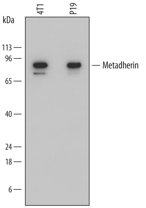
Detection of Mouse Metadherin by Western Blot.
Western blot shows lysates of 4T1 mouse breast cancer cell line and P19 mouse embryonal carcinoma cell line. PVDF membrane was probed with 1 µg/mL of Rat Anti-Mouse Metadherin Monoclonal Antibody (Catalog # MAB7180) followed by HRP-conjugated Anti-Rat IgG Secondary Antibody (Catalog # HAF005). A specific band was detected for Metadherin at approximately 80 kDa (as indicated). This experiment was conducted under reducing conditions and using Immunoblot Buffer Group 1.Applications for Mouse Metadherin Antibody
Western Blot
Sample: 4T1 mouse breast cancer cell line and P19 mouse embryonal carcinoma cell line
Reviewed Applications
Read 1 review rated 5 using MAB7180 in the following applications:
Formulation, Preparation, and Storage
Purification
Reconstitution
Formulation
Shipping
Stability & Storage
- 12 months from date of receipt, -20 to -70 °C as supplied.
- 1 month, 2 to 8 °C under sterile conditions after reconstitution.
- 6 months, -20 to -70 °C under sterile conditions after reconstitution.
Background: Metadherin
Metadherin (MTDH; also Lyric and Astrocyte-elevated gene-1/AEG1) is a unique molecule originally discovered to be upregulated in astrocytes following HIV infection. Although its predicted MW is 64 kDa, it runs anomalously at approximately 80 kDa in SDS-Page. It is widely expressed, and appears to be a component of the ER, nucleolus and inner nuclear membrane. Metadherin is reported to promote Akt/PI3 kinase activity, and suppress apoptosis-associated FOXO3A transcription. Mouse Metadherin is a type III (i.e.- a type I with no signal sequence) transmembrane protein 579 amino acids (aa) in length. It contains a luminal N-terminus (aa 1-48) with a 510 aa cytoplasmic C-terminus. There are no identifiable structural motifs, although two poly-Lys segments and two utilized Ser phosphorylation sites exist. Multiple bands at 80 kDa, 75 kDa, 50-55 kDa and 37 kDa are seen for Metadherin on SDS-Page. They may reflect the presence of at least two potential isoform variants that show 1) a deletion of aa 422-457, and 2) an Ile substitution for aa 190-579. Over aa 167-297, mouse Metadherin shares 98% and 97% aa sequence identity with rat and human Metadherin, respectively.
Alternate Names
Gene Symbol
UniProt
Additional Metadherin Products
Product Documents for Mouse Metadherin Antibody
Product Specific Notices for Mouse Metadherin Antibody
For research use only
