Mouse Rae-1 epsilon Antibody
R&D Systems, part of Bio-Techne | Catalog # MAB1135


Key Product Details
Species Reactivity
Validated:
Cited:
Applications
Validated:
Cited:
Label
Antibody Source
Product Specifications
Immunogen
Specificity
Clonality
Host
Isotype
Endotoxin Level
Scientific Data Images for Mouse Rae-1 epsilon Antibody
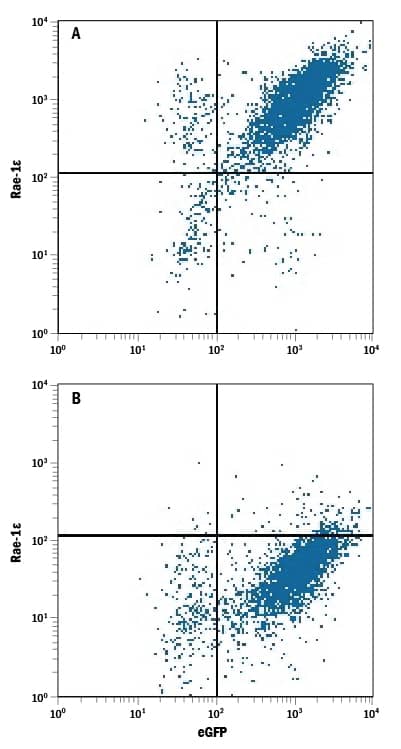
Detection of Rae‑1 epsilon in BaF3 Mouse Cell Line Transfected with Mouse Rae-1 epsilon and eGFP by Flow Cytometry.
BaF3 mouse pro-B cell line transfected with (A) mouse Rae-1e or (B) mouse Rae-1a and eGFP was stained with Rat Anti-Mouse Rae-1e Monoclonal Antibody (Catalog # MAB1135) followed by Allophycocyanin-conjugated Anti-Rat IgG Secondary Antibody (Catalog # F0113). Quadrant markers were set based on control antibody staining (Catalog # MAB006).Applications for Mouse Rae-1 epsilon Antibody
Blockade of Receptor-ligand Interaction
CyTOF-ready
Flow Cytometry
Sample: BaF3 mouse pro-B cell line transfected with mouse Rae-1 epsilon
Formulation, Preparation, and Storage
Purification
Reconstitution
Formulation
Shipping
Stability & Storage
- 12 months from date of receipt, -20 to -70 °C as supplied.
- 1 month, 2 to 8 °C under sterile conditions after reconstitution.
- 6 months, -20 to -70 °C under sterile conditions after reconstitution.
Background: Rae-1 epsilon
Rae-1 epsilon is a member of a family of cell-surface proteins that function as ligands for mouse NKG2D. Other family members are designated Rae-1 alpha, beta, gamma, and delta. Amino acid sequence identity within this family ranges from 88‑95%. The Rae-1 proteins are distantly related to MHC class I proteins, but they possess only the alpha1 and alpha2 Ig‑like domains, and they have no capacity to bind peptide or interact with beta2-microglobulin. The genes encoding these proteins are not found within the Major Histocompatibility Complex on mouse chromosome 17, but rather map to mouse chromosome 10. The Rae-1 proteins are anchored to the membrane via a GPI‑linkage. The name of this family derives from the original identification of these proteins as the product of retinoic acid early inducible transcripts. Rae-1 expression is developmentally controlled. Transcripts were observed in the brain/head region of day 10‑14 embryos but disappeared by day 18. Rae-1 transcripts were detected in several transformed cell lines but are absent from most normal adult tissues. All Rae-1 family members bind to mouse NKG2D, an activating receptor expressed on NK cells and some T cell subsets, resulting in the activation of cytolytic activity and/or cytokine production by these effector cells. Ectopic expression of Rae-1 on mouse tumor cell lines resulted in the in vivo rejection of the tumors (1‑7).
References
- Zou, Z. et al. (1996) J. Biochem (Tokyo) 119:319.
- Diefenbach, A. et al. (2000) Nature Immunol. 1:119.
- Cerwenka, A. et al. (2000) Immunity 12:721.
- Cerwenka, A. et al. (2001) Proc. Natl. Acad. Sci. USA 98:11521.
- Diefenbach, A. et al. (2001) Nature 413:165.
- Champsaur, M. et al. (2010) J. Immunol. 185:157.
- Markiewicz M. et al. (2012) Immunity 36:132.
Long Name
Alternate Names
Gene Symbol
Additional Rae-1 epsilon Products
Product Documents for Mouse Rae-1 epsilon Antibody
Product Specific Notices for Mouse Rae-1 epsilon Antibody
For research use only