Mouse/Rat Cathepsin C/DPPI Antibody
R&D Systems, part of Bio-Techne | Catalog # AF1034

Key Product Details
Validated by
Species Reactivity
Validated:
Cited:
Applications
Validated:
Cited:
Label
Antibody Source
Product Specifications
Immunogen
Asp25-Leu462
Accession # P97821
Specificity
Clonality
Host
Isotype
Scientific Data Images for Mouse/Rat Cathepsin C/DPPI Antibody
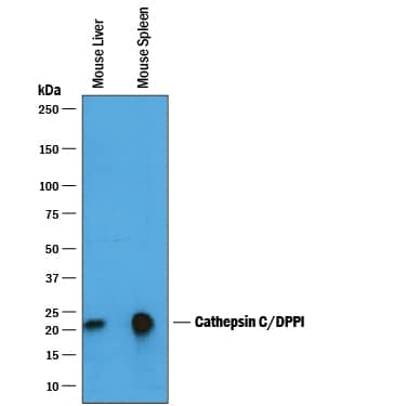
Detection of Mouse Cathepsin C/DPPI by Western Blot.
Western blot shows lysates of mouse liver tissue and mouse spleen tissue. PVDF membrane was probed with 0.25 µg/mL of Goat Anti-Mouse Cathepsin C/DPPI Antigen Affinity-purified Polyclonal Antibody (Catalog # AF1034) followed by HRP-conjugated Anti-Goat IgG Secondary Antibody (Catalog # HAF019). A specific band was detected for Cathepsin C/DPPI at approximately 20-25 kDa (as indicated). This experiment was conducted under reducing conditions and using Immunoblot Buffer Group 1.Detection of Mouse and Rat Cathepsin C/DPPI by Western Blot.
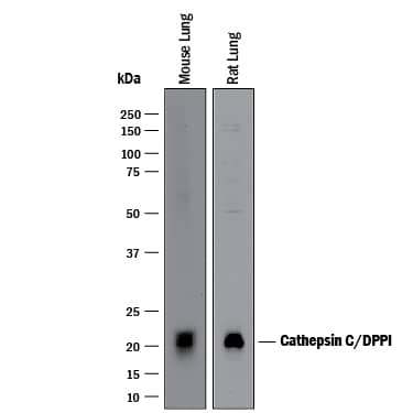
Western blot shows lysates of mouse lung tissue and rat lung tissue. PVDF membrane was probed with 1 µg/mL of Goat Anti-Mouse Cathepsin C/DPPI Antigen Affinity-purified Polyclonal Antibody (Catalog # AF1034) followed by HRP-conjugated Anti-Goat IgG Secondary Antibody (Catalog # HAF017). A specific band was detected for Cathepsin C/DPPI at approximately 20-22 kDa (as indicated). This experiment was conducted under reducing conditions and using Immunoblot Buffer Group 1.Cathepsin C/DPPI in Mouse Thymus.
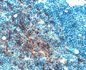
Cathepsin C/DPPI was detected in perfusion fixed frozen sections of mouse thymus using Goat Anti-Mouse Cathepsin C/ DPPI Antigen Affinity-purified Polyclonal Antibody (Catalog # AF1034) at 15 µg/mL overnight at 4 °C. Tissue was stained using the Anti-Goat HRP-DAB Cell & Tissue Staining Kit (brown; Catalog # CTS008) and counter-stained with hematoxylin (blue). Specific staining was localized to thymocytes. View our protocol for Chromogenic IHC Staining of Frozen Tissue Sections.Applications for Mouse/Rat Cathepsin C/DPPI Antibody
Immunohistochemistry
Sample: Perfusion fixed frozen section of mouse thymus
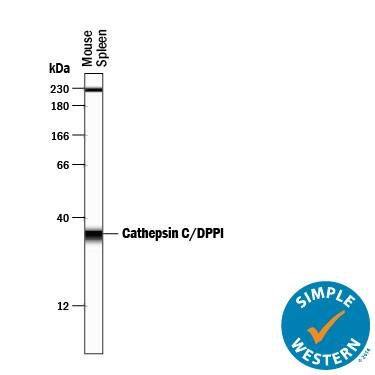
Simple Western
Sample: Mouse spleen tissue
Western Blot
Sample: Mouse liver tissue, mouse spleen tissue, mouse lung tissue, and rat lung tissue
Reviewed Applications
Read 1 review rated 3 using AF1034 in the following applications:
Formulation, Preparation, and Storage
Purification
Reconstitution
Formulation
Shipping
Stability & Storage
- 12 months from date of receipt, -20 to -70 °C as supplied.
- 1 month, 2 to 8 °C under sterile conditions after reconstitution.
- 6 months, -20 to -70 °C under sterile conditions after reconstitution.
Background: Cathepsin C/DPPI
Cathepsin C is a cysteine protease of the papain family (1). Cathepsin C sequentially removes dipeptides from the free N-termini of proteins and peptides. It has broad specificity except that it does not cleave a basic amino acid (Arg or Lys) in the N-terminal position or Pro on either side of the scissle bond. It requires halide ions for activity. The pro form contains a pro peptide and a catalytic region, which can be further processed into heavy/ alpha and light/ beta chains that are linked by a disulfide bond. It is broadly distributed. Cathepsin C plays a role in the lysosomal degradation. It also functions as a key enzyme in the activation of granule serine proteases in cytotoxic T lymphocytes and natural killer cells (granzymes A and B), mast cells (tryptase and chymase), and neutrophils (Cathepsin G and elastase) by removing their N-terminal activation dipeptides (2).
References
- Turk, B. et al. (2004) in Handbook of Proteolytic Enzymes. Barrett, et al. eds. p. 1192, Academic Press, San Diego.
- Dahl, S.W. et al. (2001) Biochemistry 40:1671.
Alternate Names
Gene Symbol
UniProt
Additional Cathepsin C/DPPI Products
Product Documents for Mouse/Rat Cathepsin C/DPPI Antibody
Product Specific Notices for Mouse/Rat Cathepsin C/DPPI Antibody
For research use only
