Rat MIS RII Antibody
R&D Systems, part of Bio-Techne | Catalog # AF1618

Key Product Details
Validated by
Species Reactivity
Validated:
Cited:
Applications
Validated:
Cited:
Label
Antibody Source
Product Specifications
Immunogen
Pro19-Pro144
Accession # Q62893
Specificity
Clonality
Host
Isotype
Scientific Data Images for Rat MIS RII Antibody
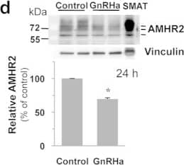
Detection of Mouse MIS RII/AMHR2 by Western Blot
GnRH down-regulates AMH receptivity in vitro in L betaT2 cells and in vivo in pituitary of female rats at pnd 18.(a) GnRH inhibits AMH signaling. L betaT2 cells were stimulated for 24 h with 2.5 μg/ml AMH, 10 nM GnRHa or with a combination of both hormones. P-Smad1/5/8 protein level was evaluated by immunoblotting and normalized with total Smad1. *P ≤ 0.05 compared with AMH-stimulated cells. (b) Combined effects of GnRH and AMH on Fshb expression. Cells were incubated with 2.5 μg/ml AMH, 10 nM GnRHa or with both hormones for 24 h. Fshb mRNA levels were analyzed by real-time qPCR and expressed as percentage of control levels. *P ≤ 0.05 ; **P ≤ 0.01 compared with control cells. (c) GnRH down-regulates Amhr2 expression. Cells were incubated with 10 nM GnRHa, 10 ng/ml activin A or 20 ng/ml BMP2 for 4 and 24 h. Amhr2 mRNA levels were determined by real-time qPCR and expressed as percentage of control cells. Results are the mean ± SEM of 8 independent experiments. *P ≤ 0.05 compared with control cells. (d) GnRH decreases AMHR2 protein level. Cells were treated for 24 h with 10 nM GnRHa and AMHR2 protein level was determined by immunoblotting after normalization with vinculin. Results are the mean ± SEM of 4 independent experiments. *P ≤ 0.05 compared with control cells. (e) GnRH down-regulates Amhr2 expression in pituitary of female rats at pnd 18. Male and female rats were injected subcutaneously at pnd 17 with 100 μl of saline solution containing or not 0.1 μg of GnRHa. Anterior pituitary Amhr2 expression was determined 24 h after injection by Taqman real time qPCR. Each value is a mean ± SEM of 8 to 14 rats. *P ≤ 0.05 compared to control rats; a, P ≤ 0.01 compared to female control rats. Image collected and cropped by CiteAb from the following publication (https://www.nature.com/articles/srep23790), licensed under a CC-BY license. Not internally tested by R&D Systems.Applications for Rat MIS RII Antibody
Blockade of Receptor-ligand Interaction
Immunohistochemistry
Sample: Perfusion fixed frozen sections of rat ovary
Western Blot
Sample: Recombinant Rat MIS RII Fc Chimera (Catalog # 1618-MR)
Formulation, Preparation, and Storage
Purification
Reconstitution
Formulation
Shipping
Stability & Storage
- 12 months from date of receipt, -20 to -70 °C as supplied.
- 1 month, 2 to 8 °C under sterile conditions after reconstitution.
- 6 months, -20 to -70 °C under sterile conditions after reconstitution.
Background: MIS RII
Müllerian inhibiting substance (MIS), also named anti-Müllerian hormone (AMH) is a tissue-specific TGF-beta superfamily growth factor. Its expression is restricted to fetal testis, plus postnatal testis and ovary (1). MIS induces Mullerian duct (female reproductive tract) regression during sexual differentiation in the male embryo and has been shown to have a regulatory role in gonads postnatally (1). Like other TGF-beta superfamily members, MIS signals via a heteromeric receptor complex consisting of a type I and a type II receptor serine/threonine kinase. Depending on the cell context, different type I receptors (including Act RIA/ALK2, BMP RIA/ALK3, and BMP RIB/ALK6) that are shared by other TGF-beta superfamily members, can be utilized for MIS signaling (1). In contrast, the type II MIS receptor (MIS RII) is unique and does not bind other TGF-beta superfamily members (1, 2). Upon ligand binding, MIS RII recruits the non-ligand binding type I receptor into the complex, resulting in phosphorylation the BMP-like signaling pathway effector proteins Smad1, Smad5 and Smad8 (1).
The gene for rat MIS RII was isolated separately by two groups working from Sertoli cell and fetal ovary cDNA libraries (3, 4). MIS RII comprises a 557 amino acid (aa) residue type I transmembrane protein with a putative 17 aa signal peptide. Mature MIS RII has a 127 aa cysteine-rich extracellular domain containing 2 potential N-glycosylation sites, a 21 aa transmembrane domain, and a 392 aa cytoplasmic region with a serine/threonine kinase domain (3, 4). Rat MIS RII shares 95% and 82% aa sequence identity with the mouse and human homologues, respectively (5). MIS RII is expressed in the mesenchymal cells surrounding the Mullerian ducts during embryonic development. Postnatally, it is expressed in uterine tissues and rodent Leydig cells, and coexpressed with MIS in the testicular Sertoli and ovarian granulosa cells (1, 6). The expression of MIS RII in the Mullerian mesenchyme is regulated by Wnt7a signaling from nearby epithelium through the canonical Wnt pathway. Wnt7a mutant mice do not express MIS RII, and do not experience Mullerian duct regression (7).
References
- Josso, N and N. diClemente (2003) Trends Endo. Met. 14:91.
- Mishna, Y. et al. (1999) Endocrinology 140:2084.
- Baarends, W. et al. (1994) Development 120:189.
- di Clement, N. et al. (1994) Mol. Endocrinol. 8:1006.
- Mishina, Y. et al. (1997) Biochem. Biophys. Res. Comm. 237:741.
- Teixeira, J. et al. (1996) Endocrinology 137:160.
- Hossain, A. and G. Saunders (2003) J. Biol. Chem. 278:26511.
Long Name
Alternate Names
Gene Symbol
UniProt
Additional MIS RII Products
Product Documents for Rat MIS RII Antibody
Product Specific Notices for Rat MIS RII Antibody
For research use only
