Rat MuSK Antibody
R&D Systems, part of Bio-Techne | Catalog # AF562


Key Product Details
Validated by
Biological Validation
Species Reactivity
Validated:
Rat
Cited:
Human, Mouse, Rat
Applications
Validated:
Neutralization, Western Blot
Cited:
Immunocytochemistry, Immunoprecipitation, Neutralization, Western Blot
Label
Unconjugated
Antibody Source
Polyclonal Goat IgG
Product Specifications
Immunogen
Mouse myeloma cell line NS0-derived recombinant rat MuSK
Specificity
Detects rat MuSK in direct ELISAs and Western blots.
Clonality
Polyclonal
Host
Goat
Isotype
IgG
Endotoxin Level
<0.10 EU per 1 μg of the antibody by the LAL method.
Scientific Data Images for Rat MuSK Antibody
Acetylcholine Receptor Clustering Mediated by MuSK and Neutralization by Rat MuSK Antibody.
Recombinant Rat Agrin (Catalog # 550-AG) induces MuSK-dependent acetylcholine receptor clustering on myotubes differentiated from the C2C12 mouse myoblast cell line in a dose-dependent manner (orange line). Acetylcholine Receptor Clustering elicited by Recombinant Rat Agrin (16 ng/mL) is neutralized (green line) by increasing concentrations of Goat Anti-Rat MuSK Antigen Affinity-purified Polyclonal Antibody (Catalog # AF562). The ND50 is typically 0.5-2 µg/mL.Detection of Human MuSK by Immunocytochemistry/ Immunofluorescence
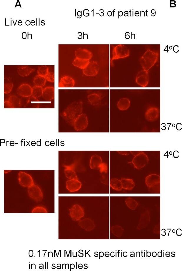
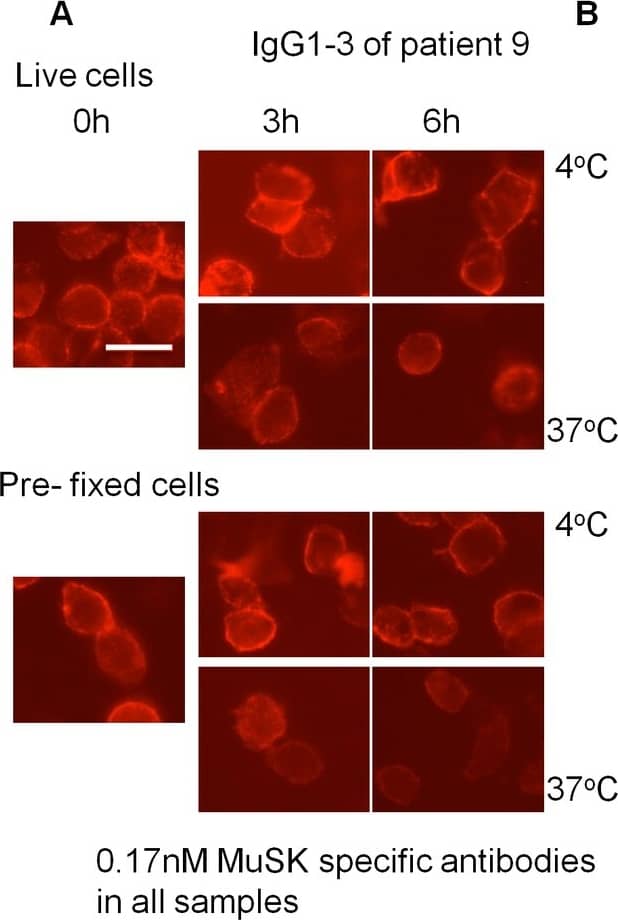
MuSK patient IgG4 or IgG1-3 do not induce endocytosis of MuSK.Patient plasma or purified IgG1-3 or IgG4 were applied at 0.17nM final concentration of MuSK antibody to HEK293 cells expressing MuSK at the cell surface. Cells were either incubated at 4°C to prevent, or at 37°C to allow, endocytosis. Patient antibody binding was visualised by addition of a secondary fluorescent anti-human antibody at the end of the experiment. (A) An example of cells that were incubated with IgG1-3 from patient 9. Robust staining was observed after 6 hours incubation at both temperatures. Scale bar=25µm. (B) Staining was scored by two individuals as described in methods, and the scores were normalised to the score at 0 hours for each condition. A slight decrease in staining was observed for cells incubated at 37°C, and pre-fixed cells also showed this reduction. (C) As a positive control for endocytosis, IgG1-3 or IgG4 was cross-linked by addition of Alexa Fluor 568-conjugated anti-human IgG prior to incubation. Example images show cells treated with patient 9 IgG4 and IgG1-3 in the presence and absence of cross-linking anti-human IgG after 6 hours incubation at 37°C. There is a clear difference when the cross-linking secondary antibody is present. Scale bar =50µm (D) Cross-linking by the secondary antibody induced internalisation of human anti-MuSK antibodies as well as MuSK-EGFP as early as 30 minutes, as observed by confocal microscopy. An example image of a cell from a confocal z-stack with orthogonal side-views is shown. The arrow shows internalised secondary Alexa Fluor 568-conjugated anti-human IgG (red) colocalised with EGFP-tagged MuSK (green). Scale bar =5µm. Image collected and cropped by CiteAb from the following open publication (https://dx.plos.org/10.1371/journal.pone.0080695), licensed under a CC-BY license. Not internally tested by R&D Systems.Detection of Human MuSK by Immunocytochemistry/ Immunofluorescence
MuSK patient IgG4 or IgG1-3 do not induce endocytosis of MuSK.Patient plasma or purified IgG1-3 or IgG4 were applied at 0.17nM final concentration of MuSK antibody to HEK293 cells expressing MuSK at the cell surface. Cells were either incubated at 4°C to prevent, or at 37°C to allow, endocytosis. Patient antibody binding was visualised by addition of a secondary fluorescent anti-human antibody at the end of the experiment. (A) An example of cells that were incubated with IgG1-3 from patient 9. Robust staining was observed after 6 hours incubation at both temperatures. Scale bar=25µm. (B) Staining was scored by two individuals as described in methods, and the scores were normalised to the score at 0 hours for each condition. A slight decrease in staining was observed for cells incubated at 37°C, and pre-fixed cells also showed this reduction. (C) As a positive control for endocytosis, IgG1-3 or IgG4 was cross-linked by addition of Alexa Fluor 568-conjugated anti-human IgG prior to incubation. Example images show cells treated with patient 9 IgG4 and IgG1-3 in the presence and absence of cross-linking anti-human IgG after 6 hours incubation at 37°C. There is a clear difference when the cross-linking secondary antibody is present. Scale bar =50µm (D) Cross-linking by the secondary antibody induced internalisation of human anti-MuSK antibodies as well as MuSK-EGFP as early as 30 minutes, as observed by confocal microscopy. An example image of a cell from a confocal z-stack with orthogonal side-views is shown. The arrow shows internalised secondary Alexa Fluor 568-conjugated anti-human IgG (red) colocalised with EGFP-tagged MuSK (green). Scale bar =5µm. Image collected and cropped by CiteAb from the following open publication (https://dx.plos.org/10.1371/journal.pone.0080695), licensed under a CC-BY license. Not internally tested by R&D Systems.Applications for Rat MuSK Antibody
Application
Recommended Usage
Western Blot
0.1 µg/mL
Sample: Recombinant Rat MuSK
Sample: Recombinant Rat MuSK
Neutralization
Measured by its ability to neutralize MuSK-dependent acetylcholine receptor clustering on myotubes differentiated from the C2C12 mouse myoblast cell line [Ferns, M.J. et al. (1993) Neuron 11:491]. The Neutralization Dose (ND50) is typically 0.5-2 µg/mL in the presence of 16 ng/mL Recombinant Rat Agrin.
Formulation, Preparation, and Storage
Purification
Antigen Affinity-purified
Reconstitution
Reconstitute at 0.2 mg/mL in sterile PBS. For liquid material, refer to CoA for concentration.
Formulation
Lyophilized from a 0.2 μm filtered solution in PBS with Trehalose. *Small pack size (SP) is supplied either lyophilized or as a 0.2 µm filtered solution in PBS.
Shipping
Lyophilized product is shipped at ambient temperature. Liquid small pack size (-SP) is shipped with polar packs. Upon receipt, store immediately at the temperature recommended below.
Stability & Storage
Use a manual defrost freezer and avoid repeated freeze-thaw cycles.
- 12 months from date of receipt, -20 to -70 °C as supplied.
- 1 month, 2 to 8 °C under sterile conditions after reconstitution.
- 6 months, -20 to -70 °C under sterile conditions after reconstitution.
Background: MuSK
Long Name
Muscle-specific Receptor Tyrosine Kinase
Alternate Names
EC 2.7.10, EC 2.7.10.1, MGC126323, MGC126324, muscle, skeletal, receptor tyrosine kinase, MuSK, skeletal receptor tyrosine-protein kinase
Gene Symbol
MUSK
Additional MuSK Products
Product Documents for Rat MuSK Antibody
Product Specific Notices for Rat MuSK Antibody
For research use only
Loading...
Loading...
Loading...
Loading...


