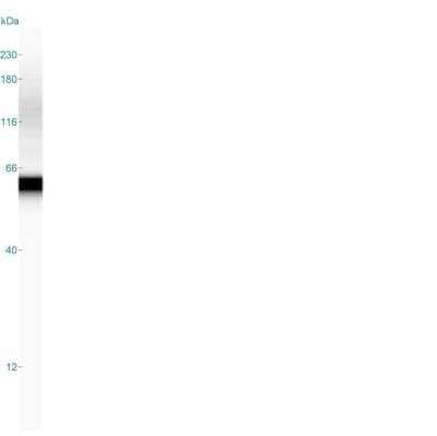
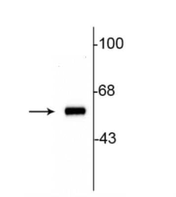
Detection of Tyrosine Hydroxylase in Rat Striatal Lysate in Western Blot
Western blot of 10 ug of rat striatal lysate showing specific immunolabeling of the ~60 kDa tyrosine hydroxylase protein.
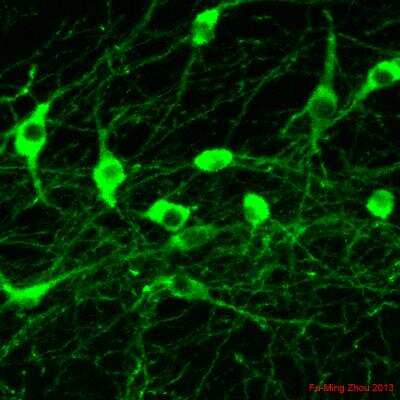
Immunohistochemical Staining of Tyrosine Hydroxylase in Frozen Mouse Tissue
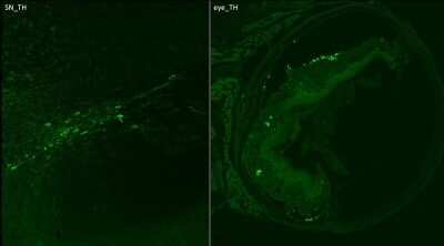
10x Magnification cryo-sections mouse: primary antibody: anti-rb TH: 1:500; o.n. secondary antibody: 1:500 dk-anti-rb DyLight488. IHC-Fr image submitted by a verified customer review.
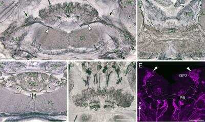
Immunohistochemical Detection of Tyrosine Hydroxylase in Cockroach Brain Tissue
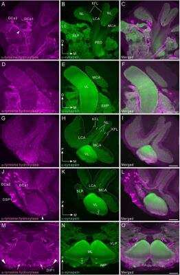
Tyrosine hydroxylase immunoreactivity in the mushroom body. All panels show a right brain hemisphere (dorsal to the top, medial to the right), except panels M-O (posterior to the top). Tyrosine hydroxylase (TH) immunoreactivity is shown in magenta (A, D, G, J, M) and synapsin immunoreactivity in green (B, E, H, K, N). Corresponding merged images in C, F, I, L, O. Scale bars = 100 um. Image collected and cropped by CiteAb from the following publication (https://dx.plos.org/10.1371/journal.pone.0160531), licensed under a CC-BY license.
Detection of Tyrosine Hydroxylase in Two Groups of Rat Adrenal Gland Extracts
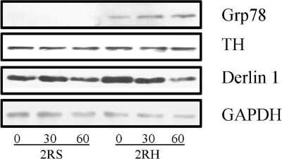
Western blot analysis confirms activation of UPR following Hotel protein lysates from the right adrenal medulla of saline (2RS) and RH groups (2RH) were subjected to Western blot analyses as described in methods. Proteins were separated by SDS-PAGE, electroblotted and the membranes were sequentially probed with antibodies specific to Grp78, Derlin1, TH and GAPDH. Similar results were obtained in two separate replicate experiments. Image collected and cropped by CiteAb from the following publication (https://dx.plos.org/10.1371/journal.pone.0172789), licensed under a CC-BY license.
Staining of Tyrosine Hydroxylase in MPC and MTT Cell Spheroids
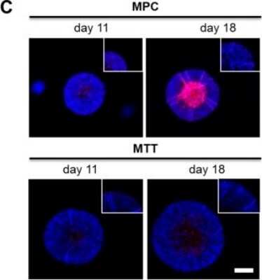
Impact of extrinsic and intrinsic hypoxia on catecholamine biosynthesis. Immunohistochemical staining showed an increased expression of Tyrosine Hydroxylase (red) in the necrotic and hypoxic core of the spheroid (DAPI, blue). Scale bar: 200 um. Image collected and cropped by CiteAb from the following publication (//pubmed.ncbi.nlm.nih.gov/31035382/) licensed under a CC-BY license.
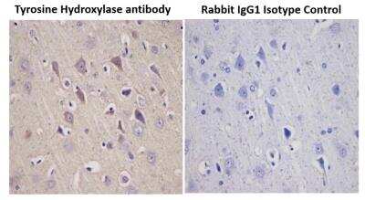
Immunohistochemical Staining of Tyrosine Hydroxylase in Paraffin Embedded Rat Brain
Immunohistochemical analysis of a formalin fixed and paraffin embedded rat brain tissue section using Tyrosine Hydroxylase antibody at 1:5000 dilution. The primary antibody binding to its antigen was detected using HRP anti-Polyvalent ready-to-use kit (Ultratek, 35368) with DAB (brown) and the sections were further counterstained using hematoxylin. Isotype control section was incubated with Rabbit IgG Isotype Control antibody and was processed under the same assay conditions. Tyrosine hydroxylase staining (brown) was observed in the test samples only and the signal was specifically localized to the neuronal cells.
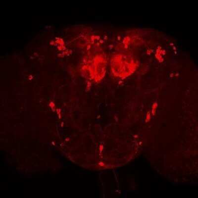
Positive Feedback on Use of Tyrosine Hydroxylase Antibody in Whole Mount Immunohistochemistry
Immunostaining of whole-mount Drosophila brains using NB300-109 at 1:500 dilution. The tyrosine hydroxylase antibody worked really well and produced a bright staining with almost no background. Data courtesy of Dr. Olga Alekseenko, Neurobiology Dept. Harvard Medical School.
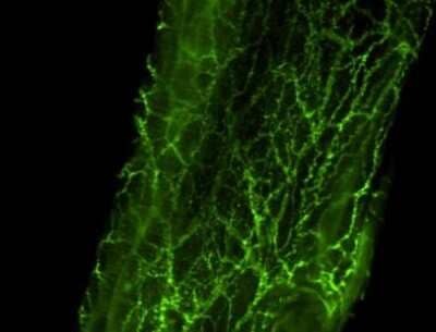
Fluorescent Staining of Tyrosine Hydroxylase in Whole Mount Rat Artery
Immunohistochemical analysis of tyrosine hydroxylase in rat mesenteric artery-whole. ICC/IF image submitted by a verified customer review.
Western Blot: Tyrosine Hydroxylase Antibody [NB300-109] -
Western blot analysis confirms activation of UPR following RH.Total protein lysates from the right adrenal medulla of saline (2RS) and RH groups (2RH) were subjected to Western blot analyses as described in methods. Proteins were separated by SDS-PAGE, electroblotted and the membranes were sequentially probed with antibodies specific to Grp78, Derlin1, TH and GAPDH. Similar results were obtained in two separate replicate experiments.
Immunocytochemistry/Immunofluorescence: Tyrosine Hydroxylase Antibody [NB300-109] -
Image showing differentiation of neurospheres as evidenced morphologically (A) and through staining by Tyrosine Hydroxylase (B) to show the ability to derive dopaminergic neurons from the rat progenitor cells.
Immunohistochemistry: Tyrosine Hydroxylase Antibody [NB300-109] -
Immunohistochemistry: Tyrosine Hydroxylase Antibody [NB300-109] - D409H GBA1 expression promotes reduction of dopaminergic fiber densities & motor deficit in the striatum of hA53T alpha-synuclein transgenic mice. a, The striatal TH-immunopositive fiber density was assessed in the striatum of the indicated genotypes at 6 months of age (n = 6 per each group). The scale bar is 250 μm. b, The optical densities of TH positive signals were quantified. c & d, Rotarod & Pole test were assessed at 6 months of age with the indicated genotypes (n = 7 per each group). e, the body weight of mice were measured with the indicated genotypes (n = 15 per each group). DATA are expressed as mean ± SEM Image collected & cropped by CiteAb from the following publication (https://actaneurocomms.biomedcentral.com/articles/10.1186/s40478-018-0538-9), licensed under a CC-BY license. Not internally tested by Novus Biologicals.
Western Blot: Tyrosine Hydroxylase Antibody [NB300-109] -
Western Blot: Tyrosine Hydroxylase Antibody [NB300-109] - Dysregulation of RNF146-PARP1 pathways in PD pathogenesis. (C) Expression of PARsylated proteins, RNF146, TH & phosphorylated alpha-synuclein (pS129-a-syn) in postmortem striatum from PD patients & age-matched control subjects (Ctrl) determined by WBs using designated antibodies. Image collected & cropped by CiteAb from following publication (https://www.oncotarget.com/lookup/doi/10.18632/oncotarget.21828), licensed under a CC-BY license. Not internally tested by Novus Biologicals.
Immunocytochemistry/ Immunofluorescence: Tyrosine Hydroxylase Antibody [NB300-109] -
Immunocytochemistry/ Immunofluorescence: Tyrosine Hydroxylase Antibody [NB300-109] - Image showing differentiation of neurospheres as evidenced morphologically (A) & through staining by Tyrosine Hydroxylase (B) to show the ability to derive dopaminergic neurons from the rat progenitor cells. Image collected & cropped by CiteAb from the following publication (https://pubmed.ncbi.nlm.nih.gov/30042680), licensed under a CC-BY license. Not internally tested by Novus Biologicals.
Western Blot: Tyrosine Hydroxylase Antibody [NB300-109] -
Western Blot: Tyrosine Hydroxylase Antibody [NB300-109] - Immunoblots of tyrosine hydroxylase in the cricket & cockroach brain.Protein extracts (8 μl & 16 μl) from the male cricket, Gryllus bimaculatus & male cockroach, Periplaneta americana were electrophoresed, transferred to a membrane & reacted with anti-tyrosine hydroxylase (TH). The anti-TH recognized a single band of protein of ~ 66 kDa (arrow heads) in both the cricket (left two lanes) & cockroach (right two lanes). M, molecular marker. Image collected & cropped by CiteAb from the following publication (https://dx.plos.org/10.1371/journal.pone.0160531), licensed under a CC-BY license. Not internally tested by Novus Biologicals.
Immunohistochemistry: Tyrosine Hydroxylase Antibody [NB300-109] -
Immunohistochemistry: Tyrosine Hydroxylase Antibody [NB300-109] - D409H GBA1 expression shortens lifespan & leads to dopaminergic degeneration in the hA53T alpha-synuclein transgenic mice. a, Survival was monitored from littermates with the following genotypes: hA53Ta-Syn/GBA1+/+ (n = 15), hA53T alpha-Syn/GBA1+/D409H(n = 16), hA53T alpha-Syn/GBA1D409H/D409H (n = 18) mice. GBA1 mutation induces the lethal phenotype & TH-positive neuronal loss in the SNpc of A53T mutant alpha-synuclein transgenic mice. b & c, The number of TH-positive neurons in the SNpc was counted using stereological analysis with the indicated genotypes at 6 months of age. The scale bar is 200 μm. DATA are expressed as mean ± SEM (n = 6 for the each group) Image collected & cropped by CiteAb from the following publication (https://actaneurocomms.biomedcentral.com/articles/10.1186/s40478-018-0538-9), licensed under a CC-BY license. Not internally tested by Novus Biologicals.
Immunocytochemistry/ Immunofluorescence: Tyrosine Hydroxylase Antibody [NB300-109] -
Immunocytochemistry/ Immunofluorescence: Tyrosine Hydroxylase Antibody [NB300-109] - Tyrosine hydroxylase immunoreactivity in the deutocerebrum & medial antennal lobe tract.Tyrosine hydroxylase (TH) immunoreactivity is shown in magenta (A, D, G, J) & synapsin immunoreactivity in green (B, E, H, K). Corresponding merged images (C, F, I, L). A-C: Frontal sections through the antennal lobe (AL). Insets indicate single optical slice (< 2 μm thickness) of the three glomeruli, which are marked by asterisks in main panel B. TH-positive fibers innervate antennal lobe glomeruli, predominantly the peripheral areas (see arrowheads in inset of A). In the macroglomerular complex (MGC), TH-positive fibers innervate exclusively the medial side (arrows in A). D-F: Horizontal sections through the antennal lobe. A single TH-positive multiglomerular projection neuron (PN) is labeled, which projects its axon toward the medial antennal lobe tract (MALT, see also panel G). G-I: Frontal sections through the medial antennal lobe tract containing several TH-positive axons (arrowheads). J-L: Frontal sections through the lobus glomerulatus (LG) & the antennal mechanosensory & motor center (AMMC). TH-immunoreactive fibers are widely distributed in the AMMC. In the lobus glomerulatus, immunoreactive fibers predominantly innervate the ventral portion. D, dorsal; M, medial; ML, medial lobe; NO, noduli; P, posterior; PB, protocerebral bridge. Scale bars = 50 μm in inset of C; 100 μm in C, F, I, L. Image collected & cropped by CiteAb from the following publication (https://dx.plos.org/10.1371/journal.pone.0160531), licensed under a CC-BY license. Not internally tested by Novus Biologicals.
Western Blot: Tyrosine Hydroxylase Antibody [NB300-109] -
Western Blot: Tyrosine Hydroxylase Antibody [NB300-109] - Specific knockdown of Hif1 alpha diminished phosphorylation of tyrosine hydroxylase & thereby reduced cellular dopamine content in PC12 cells. (A) Extrinsic hypoxia led to a time-dependent upregulation of Hif1 alpha & Hif2 alpha in PC12. (B) RNA interference using siRNA against Hif1 alpha repressed the expression of Hif1 alpha also under extrinsic hypoxia while Hif2 alpha remains unaffected. (C) Extrinsic hypoxia increased cellular dopamine content in PC12 control cells (transfection without siRNA). This effect was diminished by knockdown of Hif1 alpha. (D) Furthermore, phosphorylation of TH was decreased by knockdown of Hif1 alpha. Three independent experiments (n = 3–6). Mean ± SEM. ANOVA & Bonferroni post hoc test comparison vs. normoxia, * p < 0.05 or, ** p < 0.01, or vs. PC12 siRNA control 24 or 48 h hypoxia, # p < 0.05. Image collected & cropped by CiteAb from the following publication (https://pubmed.ncbi.nlm.nih.gov/31035382), licensed under a CC-BY license. Not internally tested by Novus Biologicals.
Immunocytochemistry/ Immunofluorescence: Tyrosine Hydroxylase Antibody [NB300-109] -
Immunocytochemistry/ Immunofluorescence: Tyrosine Hydroxylase Antibody [NB300-109] - ErbB4 protein expression in mesencephalic nuclei.The overview on the mesencephalon (B) shows dense ErbB4 expression (mAb-10, green channel) in the dopaminergic nuclei SNc & VTA that is clearly above background levels, whereas expression in the red nucleus is weaker. Higher magnification shows abundant immunoreactivity on neuropil (C–E, C'–E'). At 100x magnification, a dense network of ErbB4-positive neuropil surrounding a neuron (asterisk) is evident in the VTA (E”). The red channel shows neurofilament H-immunofluorescence in panels (B–E) & TH-immunofluorescence in (F–G). Co-immunolabeling reveals that subsets of TH-positive dopaminergic somata express ErbB4 in the VTA (F) & SNc (G); coexpressing neurons are indicated by arrowheads (F–G), whereas lack of ErbB4 signal is indicated by arrows (G). The location of the overview image within the monkey brain is indicated by a red rectangle in (A). The location of the profiles through mesencephalic nuclei is indicated by yellow boxes in (A) & by rectangles in (B). Images were obtained with a confocal microscope. Scale bar = 3 mm (B), 400 µm (C–E), 175 µm (C'–E', F–G), 70 µm (E”). Abbreviations: cerebral peduncle (cp), oculomotor nerve (3n), substantia nigra pars reticulata (SNr). Image collected & cropped by CiteAb from the following publication (https://dx.plos.org/10.1371/journal.pone.0027337), licensed under a CC0-1.0 license. Not internally tested by Novus Biologicals.
Western Blot: Tyrosine Hydroxylase Antibody [NB300-109] -
Western Blot: Tyrosine Hydroxylase Antibody [NB300-109] - Impact of extrinsic & intrinsic hypoxia on catecholamine biosynthesis. (A) Section of biosynthetic pathways of catecholamines. Squares highlighted the underlying enzymes (TH: tyrosine hydroxylase; DDC: DOPA decarboxylase; DBH: dopamine beta-hydroxylase; PNMT: phenylethanolamine N-methyltransferase). (B) Effect of extrinsic & intrinsic hypoxia on the protein expression & phosphorylation of TH at Ser40 in MTT cells. (C) Immunohistochemical staining showed an increased expression of TH (red) in the necrotic & hypoxic core of the spheroid (DAPI, blue). Scale bar: 200 µm. Catecholamine content of (D) MPC & (E) MTT cell spheroids in comparison to monolayer cultivation under normoxic or hypoxic conditions. Three independent experiments (n = 3–6). Mean ± SEM. ANOVA & Bonferroni post hoc test comparison vs. normoxia, ** p < 0.01, or vs. spheroid day 11, # p < 0.05 or, ## p < 0.01. Image collected & cropped by CiteAb from the following publication (https://pubmed.ncbi.nlm.nih.gov/31035382), licensed under a CC-BY license. Not internally tested by Novus Biologicals.
Western Blot: Tyrosine Hydroxylase Antibody [NB300-109] -
Western Blot: Tyrosine Hydroxylase Antibody [NB300-109] - Validation of protein alterations & functional studies for pathways induced by genetic & toxic stress, & reversal of PD-related alterations following S-HSP90 inhibition. a Schematic illustration of key signaling proteins & pathways activated in response to genetic stress. Western blot confirms higher activity of these pathways in PD over WT mDA neurons, & shows that PU-H71 restores these to WT levels. Mean ± SEM, n = 3 individual values from the different experiments shown as points, t-test, ***p < 0.001; **p < 0.01; *p < 0.05; ns p > 0.05. b Heatmaps of proteins significantly enriched under toxic stress (10 µM CCCP; 20 nM rotenone) & their validation by western blot (Input, total levels & S-HSP90 bait isolate, chaperome network associated). Mean ± SEM, n = 3 individual values from the different experiments shown as points, t-test, ***p < 0.001; *p < 0.05. c Western blot & quantification of rotenone stress increase in the p-TH:TH ratio & its reduction by PU-H71 (200 nM). p-ERK/ERK, cell viability control; beta-actin, protein loading control. Mean ± SEM, n = 3 individual values from the different experiments shown as points, One-way ANOVA with Tukey’s post-hoc, ***p < 0.001; **p < 0.01. d Total intracellular dopamine levels in PD mDA neurons in conditions of toxic stress & under PU-H71 rescue; Mean ± SEM, n = 4–5 individual values from the different experiments shown as points, t-test, *p < 0.05. e PU-H71 treatment of rotenone- or CCCP-stressed mDA neurons significantly increases their viability, as measured by total ATP levels; means ± SEM, n = 3–8 from independent differentiations, t-test, ***p < 0.001; **p < 0.01; *p < 0.05 Image collected & cropped by CiteAb from the following publication (https://pubmed.ncbi.nlm.nih.gov/30341316), licensed under a CC-BY license. Not internally tested by Novus Biologicals.
Immunocytochemistry/ Immunofluorescence: Tyrosine Hydroxylase Antibody [NB300-109] -
Immunocytochemistry/ Immunofluorescence: Tyrosine Hydroxylase Antibody [NB300-109] - ErbB4 protein expression in mesencephalic nuclei.The overview on the mesencephalon (B) shows dense ErbB4 expression (mAb-10, green channel) in the dopaminergic nuclei SNc & VTA that is clearly above background levels, whereas expression in the red nucleus is weaker. Higher magnification shows abundant immunoreactivity on neuropil (C–E, C'–E'). At 100x magnification, a dense network of ErbB4-positive neuropil surrounding a neuron (asterisk) is evident in the VTA (E”). The red channel shows neurofilament H-immunofluorescence in panels (B–E) & TH-immunofluorescence in (F–G). Co-immunolabeling reveals that subsets of TH-positive dopaminergic somata express ErbB4 in the VTA (F) & SNc (G); coexpressing neurons are indicated by arrowheads (F–G), whereas lack of ErbB4 signal is indicated by arrows (G). The location of the overview image within the monkey brain is indicated by a red rectangle in (A). The location of the profiles through mesencephalic nuclei is indicated by yellow boxes in (A) & by rectangles in (B). Images were obtained with a confocal microscope. Scale bar = 3 mm (B), 400 µm (C–E), 175 µm (C'–E', F–G), 70 µm (E”). Abbreviations: cerebral peduncle (cp), oculomotor nerve (3n), substantia nigra pars reticulata (SNr). Image collected & cropped by CiteAb from the following publication (https://dx.plos.org/10.1371/journal.pone.0027337), licensed under a CC0-1.0 license. Not internally tested by Novus Biologicals.
Immunocytochemistry/ Immunofluorescence: Tyrosine Hydroxylase Antibody [NB300-109] -
Immunocytochemistry/ Immunofluorescence: Tyrosine Hydroxylase Antibody [NB300-109] - Tyrosine hydroxylase immunoreactivity in mushroom body.All panels show a right brain hemisphere, except panels M-O (posterior to top). Tyrosine hydroxylase (TH) immunoreactivity shown in magenta (A, D, G, J, M) & synapsin immunoreactivity (green: B, E, H, K, N). Corresponding merged images in C, F, I, L, O. A-C: Frontal sections through lateral protocerebrum. 2 groups of TH-positive cell bodies (DCa1 & 2) labeled. DCa2 appears to send axons ventrally (arrowhead). Superior lateral protocerebrum (SLP) as well as medial & lateral calyces (MCA & LCA) innervated by TH-positive fibers. D-F: Frontal sections through vertical lobe (VL) & parts of medial calyx (MCA), which receive many fine TH-positive fibers. G-I: Horizontal sections through a tip of vertical lobe & dorsal parts of calyces. Both calyces & vertical lobe innervated by fibrous TH-positive fibers. Note TH-immunoreactive fibers exclusively innervate synapsin-positive neuropil layers (NLs) of calyx where dendrites of Kenyon cells receive synaptic inputs from antennal-lobe projection neurons. J-L: Horizontal sections through calyces, ventral to panels G-I. Here, 3clusters of TH-positive cell bodies (DCa1, DCa2, & DSP1) visible. Immunoreactive fibers mainly invade anterior layers including gamma layer ( gamma) in vertical lobe. M-O: Horizontal sections through distal portions of medial lobes (MLs), invaded by fine TH-positive fibers exhibiting moderate immunolabeling (arrows). in inferior medial protocerebrum (IMP) anterior to medial lobe, a pair of DIP1 clusters (triangles) is situated. D, dorsal; KFL, Kenyon fiber layer; LCA, lateral calyx; LO, lobula; M, medial; P, posterior; PED, pedunculus; SMP, superior medial protocerebrum. Scale bars = 100 μm. Image collected & cropped by CiteAb from following publication (https://dx.plos.org/10.1371/journal.pone.0160531), licensed under a CC-BY license. Not internally tested by Novus Biologicals.
Immunohistochemistry: Tyrosine Hydroxylase Antibody [NB300-109] -
Immunohistochemistry: Tyrosine Hydroxylase Antibody [NB300-109] - Characterization of the nigrostriatal pathway of LRRK2 conditional transgenic mice. A, Representative TH immunohistochemistry of the midbrain coronal sections of 22-month-old LRRK2 GS & GSDA transgenic & age-matched littermate controls. B, C, Stereological assessment of TH-positive (B) & Nissl-positive (C) neurons in the SNpc (control, n = 9; GS, n = 9; GS/DA, n = 9). Data are the mean number of cells per region ± SEM, n = 9 mice per group. Statistical significance was determined by two-tailed unpaired Student’s t test. D, Representative images of TH immunostaining of nerve terminals in the striatum of LRRK2 conditional transgenic mice at 22 months of age. E, Quantitation of TH immunostaining in the striatum using ImageJ software (NIH; control, n = 7; GS, n = 7; GS/DA, n = 7). Differences between groups were assessed by two-way ANOVA. Bars represent the mean ± SEM (n ≥ 5 animals/genotype). n.s., Nonsignificant. Image collected & cropped by CiteAb from the following publication (https://www.eneuro.org/lookup/doi/10.1523/ENEURO.0004-17.2017), licensed under a CC-BY license. Not internally tested by Novus Biologicals.
Immunocytochemistry/ Immunofluorescence: Tyrosine Hydroxylase Antibody [NB300-109] -
Immunocytochemistry/ Immunofluorescence: Tyrosine Hydroxylase Antibody [NB300-109] - Tyrosine hydroxylase (TH) immunoreactivity in mushroom body. Panels show a right brain hemisphere, except panels M-O (posterior top). Tyrosine hydroxylase immunoreactivity shown in magenta (A, D, G, J, M) & synapsin immunoreactivity in green (B, E, H, K, N). Corresponding merged images in C, F, I, L, O. A-C: Frontal sections through lateral protocerebrum. 2 groups of TH-positive cell bodies (DCa1 & 2) labeled. DCa2 appears to send axons ventrally (arrowhead). Superior lateral protocerebrum (SLP) as well as medial & lateral calyces (MCA & LCA) innervated by TH-positive fibers. D-F: Frontal sections through vertical lobe (VL) & parts of medial calyx (MCA), which receive many fine TH-positive fibers. G-I: Horizontal sections through tip of vertical lobe & dorsal parts of calyces. Both calyces & vertical lobe innervated by fibrous TH-positive fibers. Note TH-immunoreactive fibers exclusively innervate synapsin-positive neuropil layers (NLs) of calyx where dendrites of Kenyon cells receive synaptic inputs from antennal-lobe projection neurons. J-L: Horizontal sections through calyces, ventral to panels G-I. Here, 3clusters of TH-positive cell bodies (DCa1, DCa2, & DSP1) visible. Immunoreactive fibers mainly invade anterior layers including gamma layer ( gamma) in vertical lobe. M-O: Horizontal sections through distal portions of medial lobes (MLs), invaded by fine TH-positive fibers exhibiting moderate immunolabeling (arrows). in inferior medial protocerebrum (IMP) anterior to medial lobe, pair of DIP1 clusters (triangles) situated. D, dorsal; KFL, Kenyon fiber layer; LCA, lateral calyx; LO, lobula; M, medial; P, posterior; PED, pedunculus; SMP, superior medial protocerebrum. Scale bars = 100 μm. Image collected & cropped by CiteAb from following publication (https://dx.plos.org/10.1371/journal.pone.0160531), licensed under a CC-BY license. Not internally tested by Novus Biologicals.

![Western Blot: Tyrosine Hydroxylase Antibody [NB300-109] - Tyrosine Hydroxylase Antibody](https://resources.bio-techne.com/images/products/nb300-109_rabbit-polyclonal-tyrosine-hydroxylase-antibody-271220231253362.jpg)
![Immunocytochemistry/Immunofluorescence: Tyrosine Hydroxylase Antibody [NB300-109] - Tyrosine Hydroxylase Antibody](https://resources.bio-techne.com/images/products/nb300-109_rabbit-polyclonal-tyrosine-hydroxylase-antibody-271220231253335.jpg)
![Immunohistochemistry: Tyrosine Hydroxylase Antibody [NB300-109] - Tyrosine Hydroxylase Antibody](https://resources.bio-techne.com/images/products/nb300-109_rabbit-polyclonal-tyrosine-hydroxylase-antibody-310202415371995.jpg)
![Western Blot: Tyrosine Hydroxylase Antibody [NB300-109] - Tyrosine Hydroxylase Antibody](https://resources.bio-techne.com/images/products/nb300-109_rabbit-polyclonal-tyrosine-hydroxylase-antibody-310202415293344.jpg)
![Immunocytochemistry/ Immunofluorescence: Tyrosine Hydroxylase Antibody [NB300-109] - Tyrosine Hydroxylase Antibody](https://resources.bio-techne.com/images/products/nb300-109_rabbit-polyclonal-tyrosine-hydroxylase-antibody-310202415392574.jpg)
![Western Blot: Tyrosine Hydroxylase Antibody [NB300-109] - Tyrosine Hydroxylase Antibody](https://resources.bio-techne.com/images/products/nb300-109_rabbit-polyclonal-tyrosine-hydroxylase-antibody-31020241535660.jpg)
![Immunohistochemistry: Tyrosine Hydroxylase Antibody [NB300-109] - Tyrosine Hydroxylase Antibody](https://resources.bio-techne.com/images/products/nb300-109_rabbit-polyclonal-tyrosine-hydroxylase-antibody-310202415395928.jpg)
![Immunocytochemistry/ Immunofluorescence: Tyrosine Hydroxylase Antibody [NB300-109] - Tyrosine Hydroxylase Antibody](https://resources.bio-techne.com/images/products/nb300-109_rabbit-polyclonal-tyrosine-hydroxylase-antibody-310202416212328.jpg)
![Western Blot: Tyrosine Hydroxylase Antibody [NB300-109] - Tyrosine Hydroxylase Antibody](https://resources.bio-techne.com/images/products/nb300-109_rabbit-polyclonal-tyrosine-hydroxylase-antibody-310202416205183.jpg)
![Immunocytochemistry/ Immunofluorescence: Tyrosine Hydroxylase Antibody [NB300-109] - Tyrosine Hydroxylase Antibody](https://resources.bio-techne.com/images/products/nb300-109_rabbit-polyclonal-tyrosine-hydroxylase-antibody-310202416205171.jpg)
![Western Blot: Tyrosine Hydroxylase Antibody [NB300-109] - Tyrosine Hydroxylase Antibody](https://resources.bio-techne.com/images/products/nb300-109_rabbit-polyclonal-tyrosine-hydroxylase-antibody-31020241621826.jpg)
![Western Blot: Tyrosine Hydroxylase Antibody [NB300-109] - Tyrosine Hydroxylase Antibody](https://resources.bio-techne.com/images/products/nb300-109_rabbit-polyclonal-tyrosine-hydroxylase-antibody-31020241620377.jpg)
![Immunocytochemistry/ Immunofluorescence: Tyrosine Hydroxylase Antibody [NB300-109] - Tyrosine Hydroxylase Antibody](https://resources.bio-techne.com/images/products/nb300-109_rabbit-polyclonal-tyrosine-hydroxylase-antibody-310202416212334.jpg)
![Immunocytochemistry/ Immunofluorescence: Tyrosine Hydroxylase Antibody [NB300-109] - Tyrosine Hydroxylase Antibody](https://resources.bio-techne.com/images/products/nb300-109_rabbit-polyclonal-tyrosine-hydroxylase-antibody-310202416212326.jpg)
![Immunohistochemistry: Tyrosine Hydroxylase Antibody [NB300-109] - Tyrosine Hydroxylase Antibody](https://resources.bio-techne.com/images/products/nb300-109_rabbit-polyclonal-tyrosine-hydroxylase-antibody-310202416205143.jpg)
![Immunocytochemistry/ Immunofluorescence: Tyrosine Hydroxylase Antibody [NB300-109] - Tyrosine Hydroxylase Antibody](https://resources.bio-techne.com/images/products/nb300-109_rabbit-polyclonal-tyrosine-hydroxylase-antibody-310202416205113.jpg)