Recombinant Mouse Semaphorin 4A mFc Chimera Protein, CF
R&D Systems, part of Bio-Techne | Catalog # 11096-S4

Key Product Details
Source
NS0
Accession #
Structure / Form
Disulfide-linked homodimer
Conjugate
Unconjugated
Applications
Bioactivity
Product Specifications
Source
Mouse myeloma cell line, NS0-derived mouse Semaphorin 4A protein
| Mouse Sema 4A (Thr33-Phe683) Accession # Q62178.2 |
IEGRMDP | Mouse IgG2a (Glu98-Lys330) |
| N-terminus | C-terminus |
Purity
>90%, by SDS-PAGE visualized with Silver Staining and quantitative densitometry by Coomassie® Blue Staining.
Endotoxin Level
<0.10 EU per 1 μg of the protein by the LAL method.
N-terminal Sequence Analysis
Thr33
Predicted Molecular Mass
99 kDa
SDS-PAGE
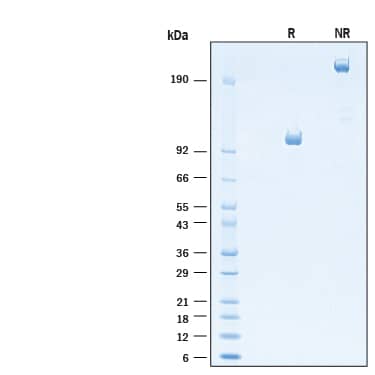
90-110 kDa, under reducing conditions.
Activity
Measured by its binding ability in a functional ELISA.
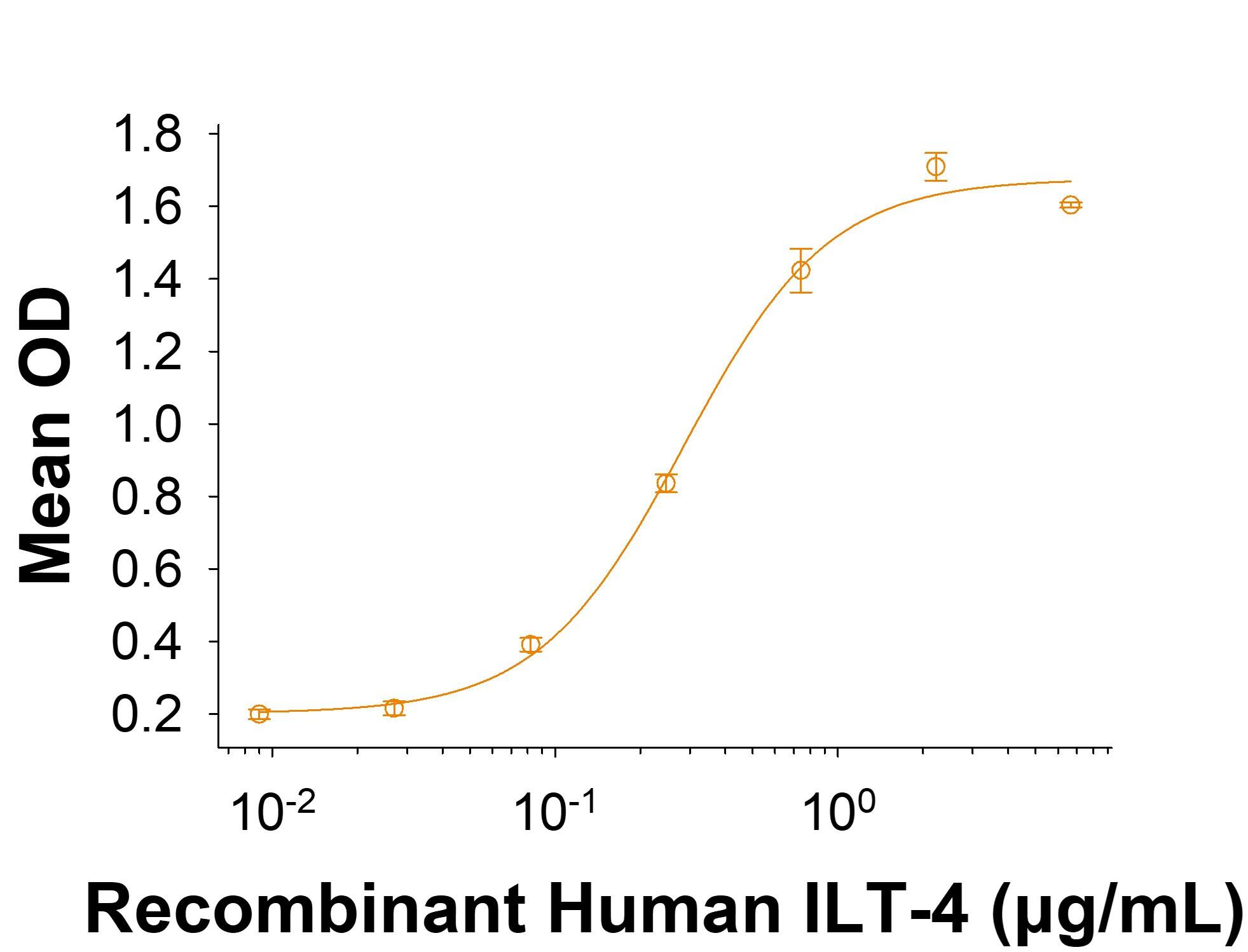
When Recombinant Mouse Semaphorin 4A mFc Chimera (Catalog # 11096-S4) is immobilized at 2.0 µg/mL (100 µL/well), the concentration of Recombinant Human LILRB2/CD85d/ILT4 Fc Chimera Protein (Catalog # 2078-T4) that produces 50% of the optimal binding response is 0.100-0.600 μg/mL
When Recombinant Mouse Semaphorin 4A mFc Chimera (Catalog # 11096-S4) is immobilized at 2.0 µg/mL (100 µL/well), the concentration of Recombinant Human LILRB2/CD85d/ILT4 Fc Chimera Protein (Catalog # 2078-T4) that produces 50% of the optimal binding response is 0.100-0.600 μg/mL
Scientific Data Images for Recombinant Mouse Semaphorin 4A mFc Chimera Protein, CF
Recombinant Mouse Semaphorin 4A mFc Chimera Protein Bioactivity.
When Recombinant Mouse Semaphorin 4A mFc Chimera Protein (Catalog #11096-S4) is immobilized at 2.0 µg/mL (100 µL/well), the concentration of Recombinant Human LILRB2/CD85d/ILT4 Fc Chimera Protein (2078-T4) that produces 50% of the optimal binding response is 0.100-0.600 μg/mLRecombinant Mouse Semaphorin 4A mFc Chimera Protein SDS-PAGE.
2 μg/lane of Recombinant Mouse Semaphorin 4A mFc Chimera Protein (Catalog # 11096-S4) was resolved with SDS-PAGE under reducing (R) and non-reducing (NR) conditions and visualized by Coomassie® Blue staining, showing bands at 90-110 kDa and 180-220 kDa, respectively.Formulation, Preparation and Storage
11096-S4
| Formulation | Lyophilized from a 0.2 μm filtered solution in PBS with Trehalose. |
| Reconstitution | Reconstitute at 250 μg/mL in PBS. |
| Shipping | The product is shipped at ambient temperature. Upon receipt, store it immediately at the temperature recommended below. |
| Stability & Storage | Use a manual defrost freezer and avoid repeated freeze-thaw cycles.
|
Background: Semaphorin 4A
References
- Kumanogoh, A. et al. (2003) J. Cell Sci. 116:3463.
- Swissprot Accession # Q9H3S1.
- Entrez Accession # CAI15528, CAI15529, CAI15531, CAI15532, CAI15533 and EAW52993.
- Kumanogoh, A. et al. (2002) Nature 419:629.
- Lu, N. et al. (2018) Nature Commun. 9:742.
- Kumanogoh, A. et al. (2005) Immunity 22:305.
- Yukawa, K. et al. (2005) Int. J. Mol. Med. 16:115.
- Toyofuku, T. et al. (2007) EMBO J. 26:1373.
- Rice, D.S. et al. (2004) Invest. Ophthalmol. Vis. Sci. 45:2767.
- Abid, A. et al. (2007) J. Med. Genet. 43:378.
Alternate Names
CORD10, SEMA4A, SEMAB, SEMB
Gene Symbol
SEMA4A
UniProt
Additional Semaphorin 4A Products
Product Documents for Recombinant Mouse Semaphorin 4A mFc Chimera Protein, CF
Product Specific Notices for Recombinant Mouse Semaphorin 4A mFc Chimera Protein, CF
For research use only
Loading...
Loading...
Loading...

